
















©The Author(s) 2025.
World J Stem Cells. Oct 26, 2025; 17(10): 109284
Published online Oct 26, 2025. doi: 10.4252/wjsc.v17.i10.109284
Published online Oct 26, 2025. doi: 10.4252/wjsc.v17.i10.109284
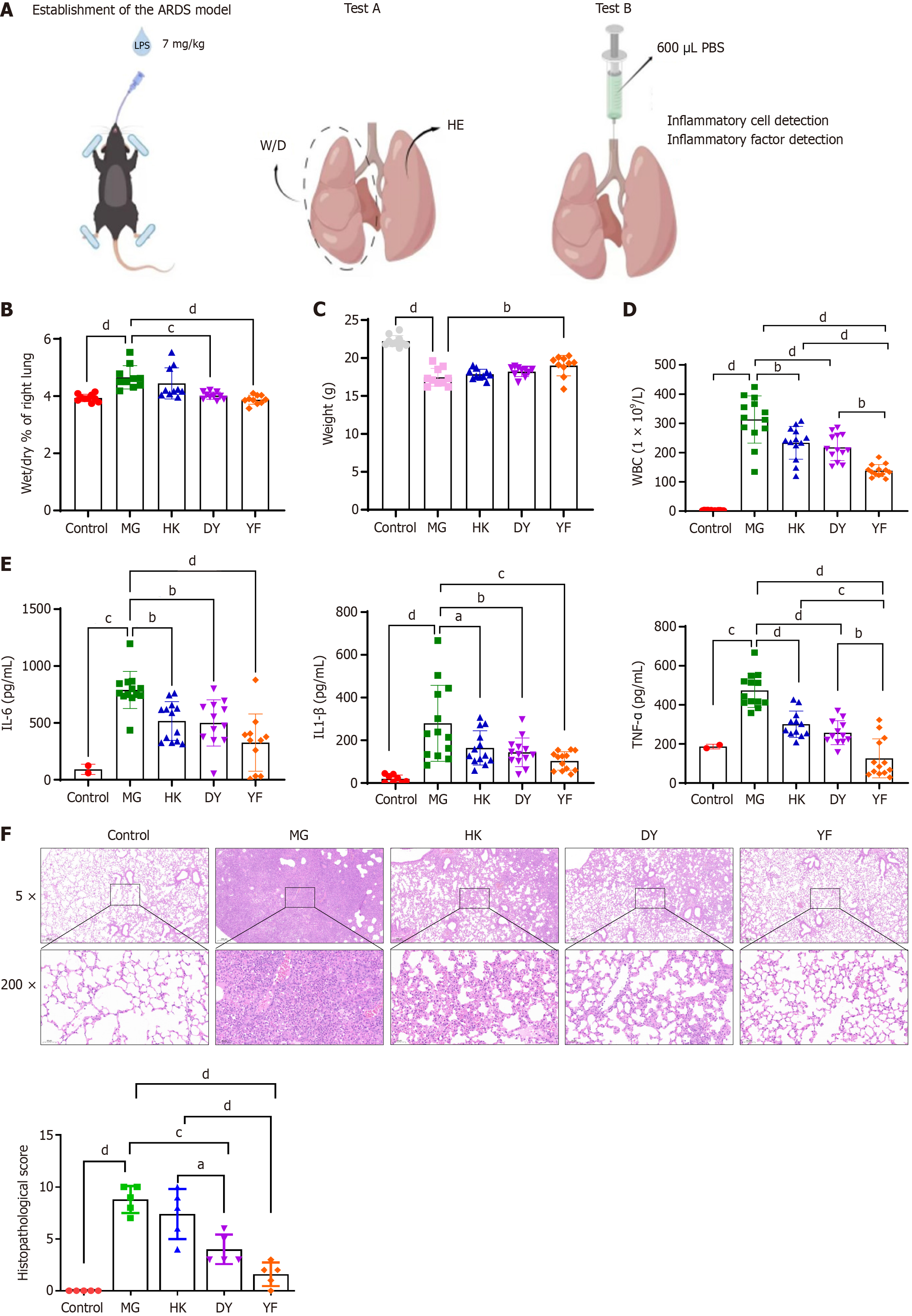
Figure 1 Effects of mesenchymal stromal cells in various media on acute respiratory distress syndrome.
A: Diagram illustrating the acute respiratory distress syndrome study design; B and C: Comparison of the wet-dry ratio (B) and body weight (C) of lung tissue from different groups; D and E: Cytokine levels in bronchoalveolar lavage fluid were measured at the experimental endpoint; F: Pathological changes and lung injury scores of the lung tissue in each group. The data are presented as means ± SD. Statistical analysis between groups was performed via two-sided Student’s t test, and within groups, ANOVA was used for normally distributed data. n = 10, aP < 0.05, bP < 0.01, cP < 0.001, dP < 0.0001. ARDS: Acute respiratory distress syndrome; W/D: Wet-dry ratio; HE: Hematoxylin and eosin; PBS: Phosphate buffered saline; MG: Model control (vehicle administered via caudal vein injection after establishing the acute respiratory distress syndrome model; DY: Mesenchymal stromal cells cultured in medium from Dakewe Biotech Co., administered via caudal vein injection after establishing the acute respiratory distress syndrome model; YF: Mesenchymal stromal cells cultured in medium from Shandong Qilu Cell Therapy Engineering Technology Co. Ltd., administered via caudal vein injection under the acute respiratory distress syndrome model; HK: Mesenchymal stromal cells cultured in medium from Cytoniche, administered via caudal vein injection in the acute respiratory distress syndrome model; WBC: White blood cell; IL: Interleukin; TNF: Tumor necrosis factor.
- Citation: Zhang YL, Wen DK, Wang SN, Tan Y, Ma HR. Melanoma cell adhesion molecule-positive mesenchymal stromal cells alleviate acute respiratory distress syndrome via nuclear factor kappa-B-mediated paracrine regulation. World J Stem Cells 2025; 17(10): 109284
- URL: https://www.wjgnet.com/1948-0210/full/v17/i10/109284.htm
- DOI: https://dx.doi.org/10.4252/wjsc.v17.i10.109284