Copyright
©The Author(s) 2024.
World J Stem Cells. Apr 26, 2024; 16(4): 410-433
Published online Apr 26, 2024. doi: 10.4252/wjsc.v16.i4.410
Published online Apr 26, 2024. doi: 10.4252/wjsc.v16.i4.410
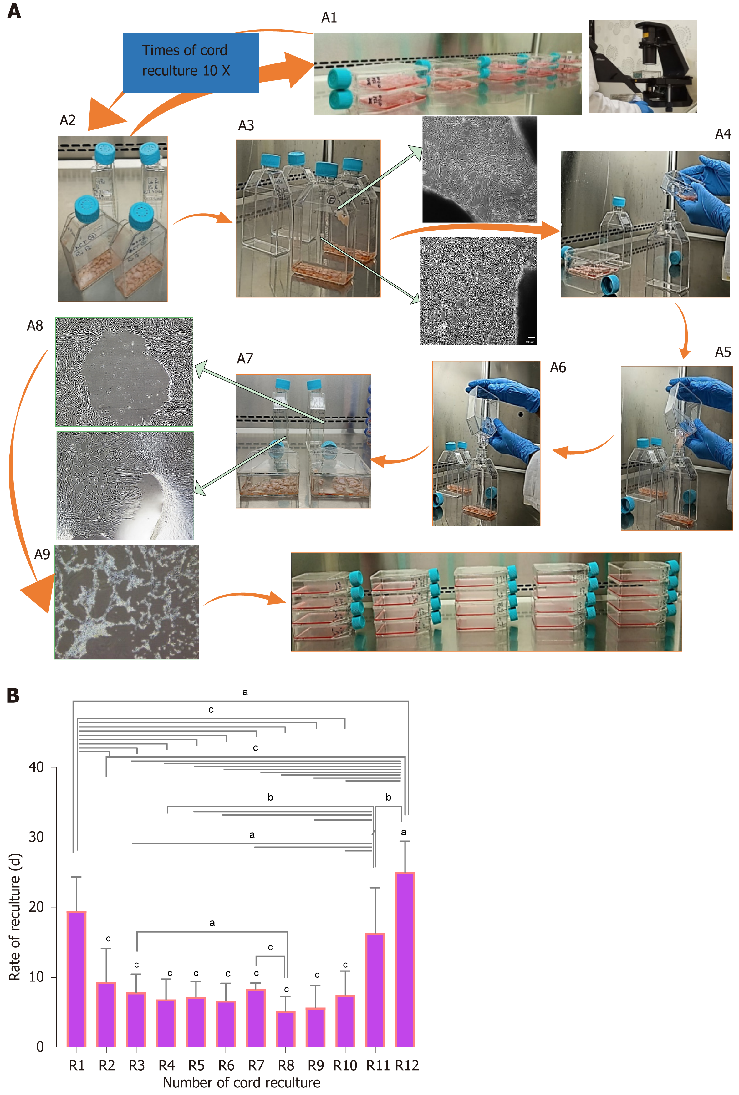
Figure 3 Effect of reculturing human umbilical cord for mesenchymal stem cell expansion and reculturing rate.
A: Novel design of recultured human umbilical cord (hUC)-mesenchymal stem cells scaling method; A1: Microscopic observation of explant containing flask at respective recultured days mentioned in Figure 2; A2: Selection of flasks that occupied maximal 80%-90 % confluent colonies of mesenchymal stem cells (MSCs) releasing from the explant; A3: Microscopic observation of explant releasing MSCs; A4: Maintaining sterile culture conditions marking new flasks as subsequent recultured number and gently tap the explant-containing flask to propel all surface sticky cord pieces towards the canted neck of the flask; A5: Inclined explant containing MSC confluent flask at an angular position of 45-90 degree over the new flask with marked successive recultured number aseptically; A6: Transfer all the explant pieces into the new flask; A7 and A8: Microscopic representation of confluent hUC-MSCs after removing the explant; A9: Sub-culturing of explant removed adherent MSCs showing cellular detachment by trypsinization; A10: Scaled MSCs for experimentation; B: The graphical representation of recultured hUC-MSC: The two-way ANOVA with multiple comparisons of recultured hUC between R1-R12 times. The recultured numbers 1-10 showed a significant decrease in time to recultured hUC, hence increased MSC expansion. At R11 and R12 a significant increase in time was observed to get MSCs. aP < 0.05, bP < 0.01, and cP < 0.001.
- Citation: Rajput SN, Naeem BK, Ali A, Salim A, Khan I. Expansion of human umbilical cord derived mesenchymal stem cells in regenerative medicine. World J Stem Cells 2024; 16(4): 410-433
- URL: https://www.wjgnet.com/1948-0210/full/v16/i4/410.htm
- DOI: https://dx.doi.org/10.4252/wjsc.v16.i4.410


