Copyright
©The Author(s) 2023.
World J Stem Cells. Nov 26, 2023; 15(11): 999-1016
Published online Nov 26, 2023. doi: 10.4252/wjsc.v15.i11.999
Published online Nov 26, 2023. doi: 10.4252/wjsc.v15.i11.999
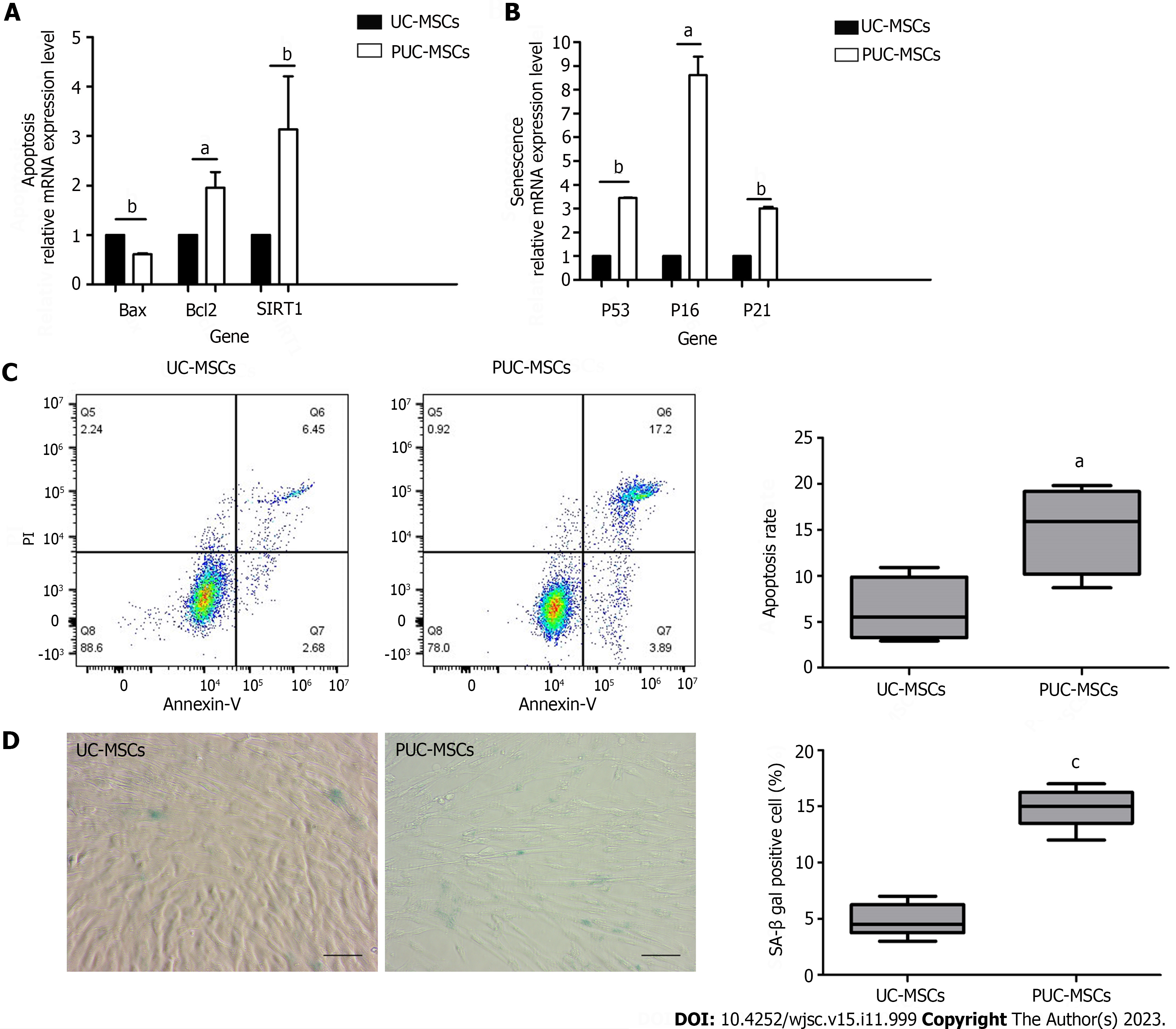
Figure 4 Effects of pretreatment on apoptosis and senescence in mesenchymal stem cells.
A: Apoptosis-related gene analysis showed that the expression of B-cell lymphoma 2 (BCL-2)-associated protein (Bax) was decreased and the expression of Bcl2 and silencing information regulator 2-related enzyme 1 (SIRT1) was increased after hypoxia and inflammatory factor pretreatment; B: Expression of P53, P16 and P21 was upregulated after hypoxia and inflammatory factor pretreatment; C: Umbilical cord mesenchymal stem cells (UC-MSCs) and primed UC-MSCs (PUC-MSCs) were stained with Annexin V-FITC apoptosis assay kit reagents and apoptotic cells were detected by flow cytometry. The results showed that the apoptosis index of PUC-MSCs increased; D: Representative images of β-galactosidase (SA-β-gal) staining and quantitative analysis of positive SA-β-gal staining. Compared with that of the UC-MSCs, the number of SA-β-gal-positive PUC-MSCs was significantly increased; Bar = 100 μm. aP < 0.05; bP < 0.01; cP < 0.001.
- Citation: Li H, Ji XQ, Zhang SM, Bi RH. Hypoxia and inflammatory factor preconditioning enhances the immunosuppressive properties of human umbilical cord mesenchymal stem cells. World J Stem Cells 2023; 15(11): 999-1016
- URL: https://www.wjgnet.com/1948-0210/full/v15/i11/999.htm
- DOI: https://dx.doi.org/10.4252/wjsc.v15.i11.999


