Copyright
©The Author(s) 2023.
World J Stem Cells. Nov 26, 2023; 15(11): 999-1016
Published online Nov 26, 2023. doi: 10.4252/wjsc.v15.i11.999
Published online Nov 26, 2023. doi: 10.4252/wjsc.v15.i11.999
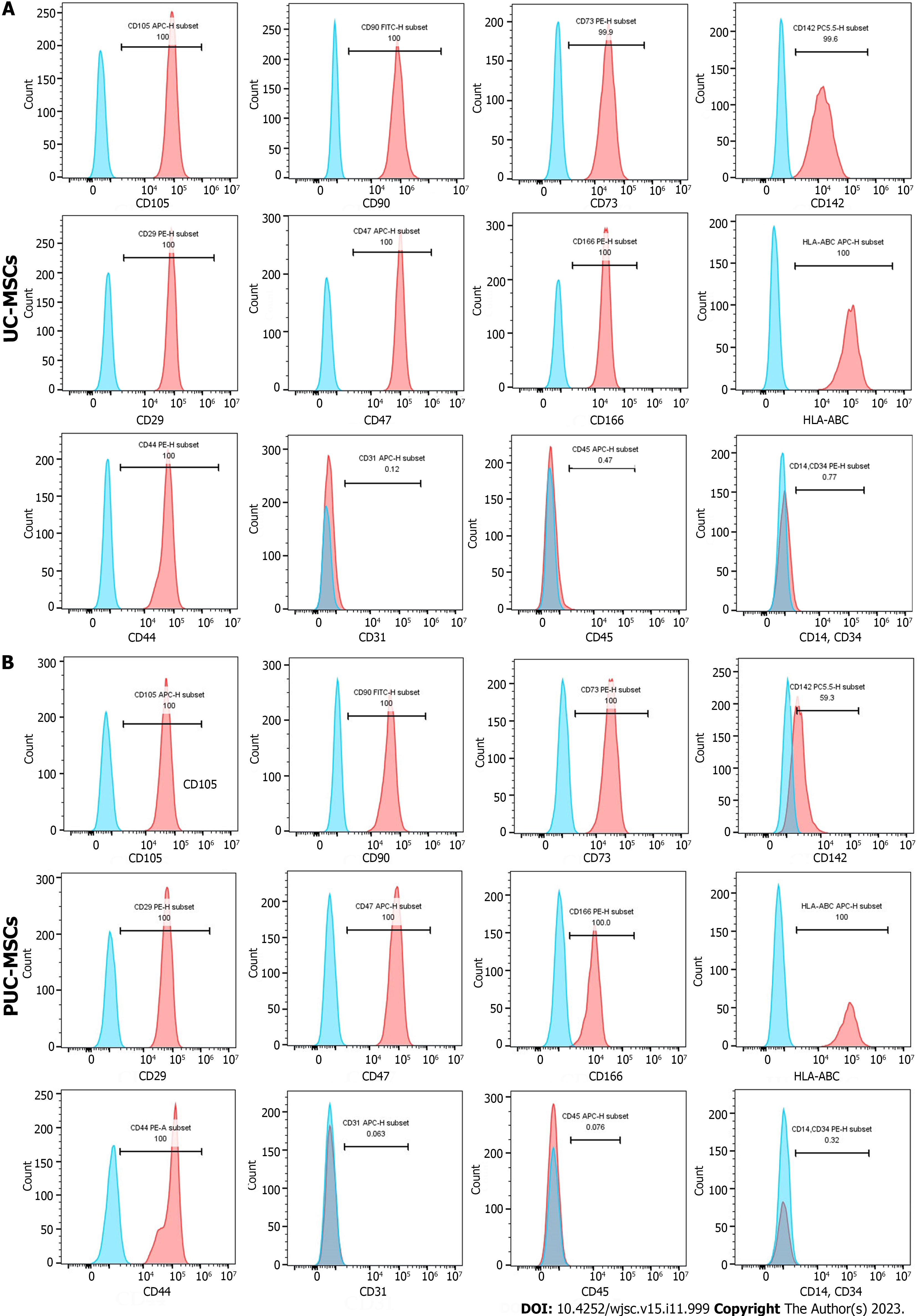
Figure 2 Phenotypes of mesenchymal stem cells were detected by flow cytometry.
A: Cell surface markers of umbilical cord mesenchymal stem cells (UC-MSCs); B: Cell surface markers of primed umbilical cord mesenchymal stem cells (PUC-MSCs). The signals from the unstained control cells are shown in blue in the histogram, and the signals from stained cells are shown in red in the histogram. HLA: Human leukocyte antigen.
- Citation: Li H, Ji XQ, Zhang SM, Bi RH. Hypoxia and inflammatory factor preconditioning enhances the immunosuppressive properties of human umbilical cord mesenchymal stem cells. World J Stem Cells 2023; 15(11): 999-1016
- URL: https://www.wjgnet.com/1948-0210/full/v15/i11/999.htm
- DOI: https://dx.doi.org/10.4252/wjsc.v15.i11.999


