Copyright
©The Author(s) 2022.
World J Stem Cells. Jul 26, 2022; 14(7): 527-538
Published online Jul 26, 2022. doi: 10.4252/wjsc.v14.i7.527
Published online Jul 26, 2022. doi: 10.4252/wjsc.v14.i7.527
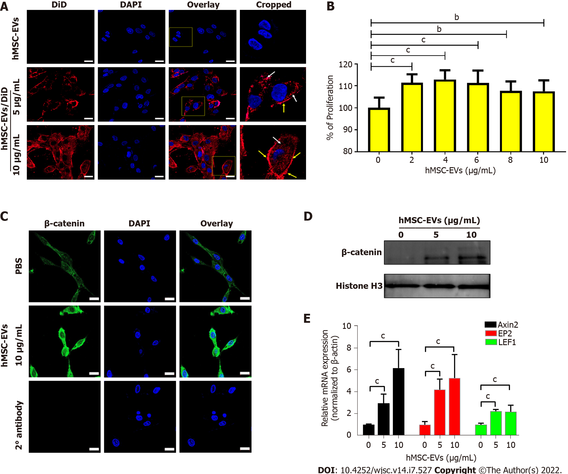
Figure 2 Interaction of human mesenchymal stem cell-derived extracellular vesicles with dermal papillae cells leads to cell proliferation and activation of Wnt/β-catenin signaling.
A: Dermal papillae (DP) cells incubated for 2 h with non-labeled human mesenchymal stem cell-derived extracellular vesicles (hMSC-EVs) (10 μg/mL) and DiD-labeled hMSC-EVs (5 and 10 μg/mL; hMSC-EVs/DiD) (scale bar: 20 μm); B: DP cell proliferation was determined using a CCK8 assay 24 h after treatment with 0–10 μg hMSC-EVs (n = 5); C: β-catenin immunofluorescence assay in DP cells after 24 h of treatment with hMSC-EVs (10 μg/mL) (scale bar: 20 μm); D: The levels of β-catenin in the nuclear fraction of DP cells treated with hMSC-EVs (5 and 10 μg/mL) with histone H3 used as a loading control for nuclear fraction; E: Quantitative real-time polymerase chain reaction results of mRNA expression of Axin2, EP2 and LEF1 in DP cells treated with hMSC-EVs (5 and 10 μg/mL) for 24 h (n = 3). The values obtained from experiments are shown mean ± SD (bP < 0.01; cP < 0.001. Student’s t-test was used for comparison). hMSC-EVs: Human mesenchymal stem cell-derived extracellular vesicles; DP: Dermal papillae.
- Citation: Rajendran RL, Gangadaran P, Kwack MH, Oh JM, Hong CM, Sung YK, Lee J, Ahn BC. Application of extracellular vesicles from mesenchymal stem cells promotes hair growth by regulating human dermal cells and follicles. World J Stem Cells 2022; 14(7): 527-538
- URL: https://www.wjgnet.com/1948-0210/full/v14/i7/527.htm
- DOI: https://dx.doi.org/10.4252/wjsc.v14.i7.527


