Copyright
©The Author(s) 2022.
World J Stem Cells. May 26, 2022; 14(5): 330-346
Published online May 26, 2022. doi: 10.4252/wjsc.v14.i5.330
Published online May 26, 2022. doi: 10.4252/wjsc.v14.i5.330
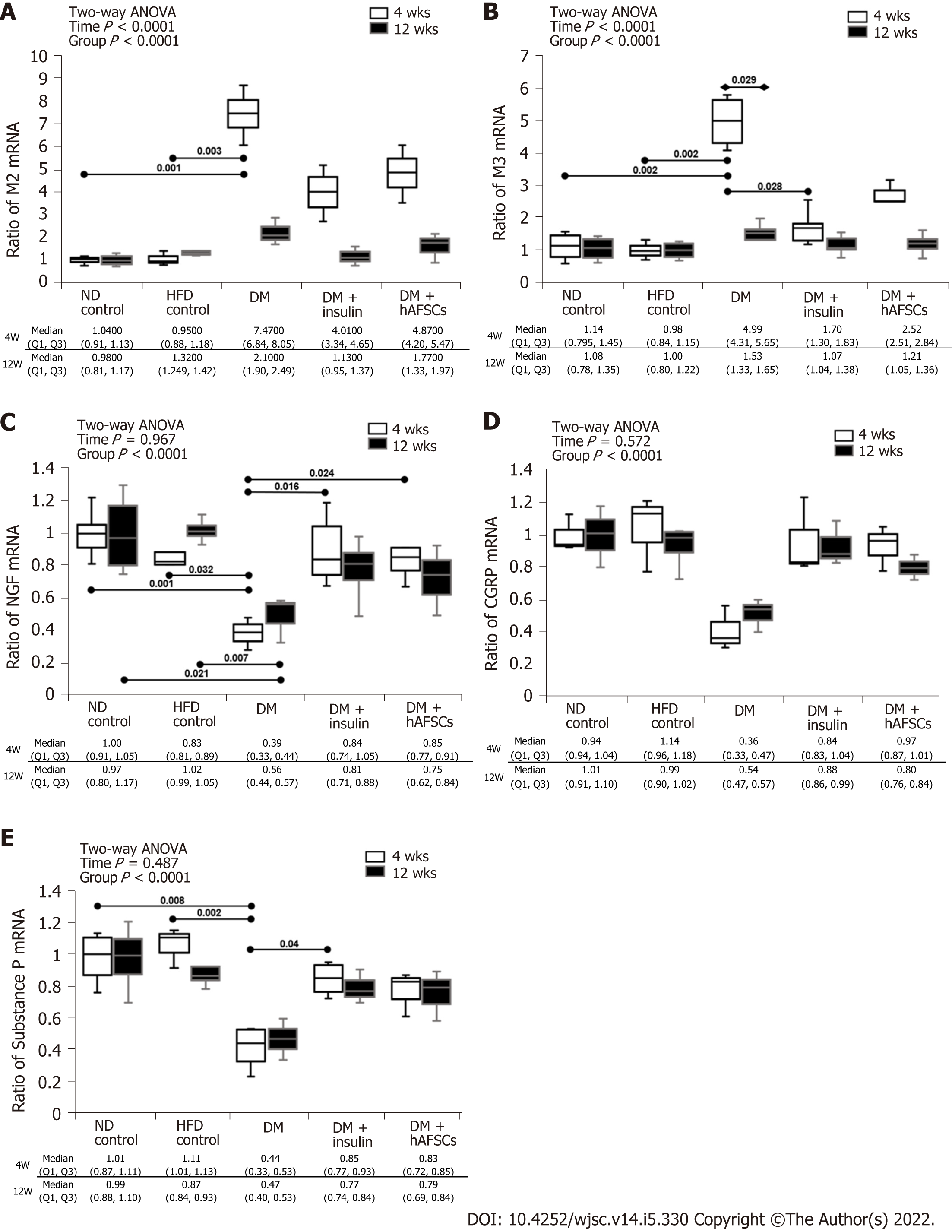
Figure 3 Relative mRNA expression.
In diabetes (DM) rats, the M3 mRNA expression was increased at 4 wk compared with 12 wk and the M2/M3 mRNA expression at 4 wk was increased compared with normal diet (ND)/high-fat diet (HFD) control groups. The M3 mRNA recovered at 4 wk after insulin treatment. The DM rats had significantly decreased mRNA expression of nerve growth factor (NGF) at 4 and 12 wk and substance P at 4 wk compared with ND/HFD controls. Insulin treatment can recover the mRNA expression of NGF and substance P, and hAFSCs treatment can recover the mRNA expression of NGF at 4 wk after DM induction. n = 6. Statistics: Median with first and third quartile (Q1, Q3) for continuous variables. Two-way analysis of variance was used for initial analysis. Kruskal-Wallis test with post hoc Bonferroni test for intergroup analysis. Mann-Whitney U test for the comparison between 4 wk and 12 wk. P values are shown in each comparison. A: M2-muscarinic receptor; B: M3-muscarinic receptor; C: Nerve growth factor; D: Calcitonin gene-related peptide; E: Substance P in the bladder of rats of different groups. ND: Normal diet; DM: Diabetes mellitus; hAFSCs: Human amniotic fluid stem cells; HFD: High-fat diet.
- Citation: Liang CC, Shaw SW, Huang YH, Lee TH. Human amniotic fluid stem cell therapy can help regain bladder function in type 2 diabetic rats. World J Stem Cells 2022; 14(5): 330-346
- URL: https://www.wjgnet.com/1948-0210/full/v14/i5/330.htm
- DOI: https://dx.doi.org/10.4252/wjsc.v14.i5.330


