Copyright
©The Author(s) 2022.
World J Stem Cells. Apr 26, 2022; 14(4): 303-309
Published online Apr 26, 2022. doi: 10.4252/wjsc.v14.i4.303
Published online Apr 26, 2022. doi: 10.4252/wjsc.v14.i4.303
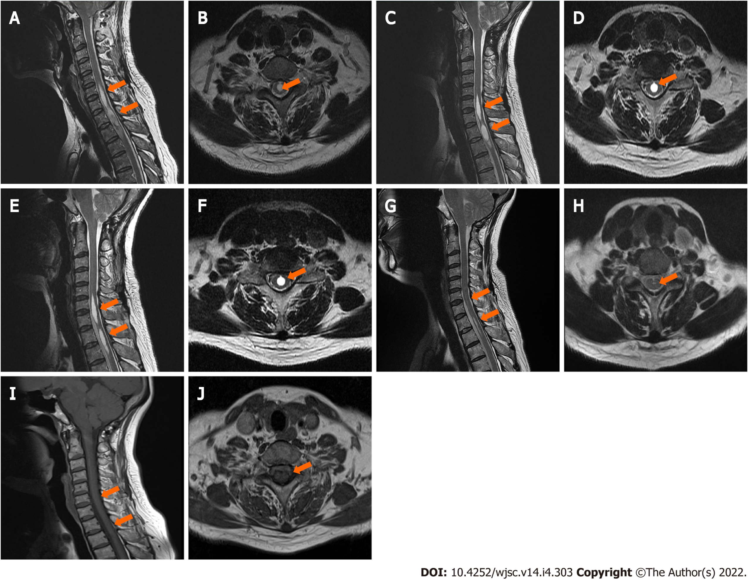
Figure 1 Imaging analyses before and after stem cell treatment.
A-F: Magnetic resonance imaging scans of the patient before stem cell treatment, from November 26, 2010 (A: Sagittal; B: Transverse), August 29, 2011 (C: Sagittal; D: Transverse) and August 21, 2012 (E: Sagittal; F: Transverse); G-J: Magnetic resonance imaging of the patient after stem cell treatment, from July 18, 2018 (G: Sagittal; H: Transverse) and August 31, 2020 (I: Sagittal; J: Transverse). Orange arrows indicate where the cavity is or was.
- Citation: Ahn H, Lee SY, Jung WJ, Lee KH. Treatment of syringomyelia using uncultured umbilical cord mesenchymal stem cells: A case report and review of literature. World J Stem Cells 2022; 14(4): 303-309
- URL: https://www.wjgnet.com/1948-0210/full/v14/i4/303.htm
- DOI: https://dx.doi.org/10.4252/wjsc.v14.i4.303


