Copyright
©The Author(s) 2022.
World J Stem Cells. Mar 26, 2022; 14(3): 245-263
Published online Mar 26, 2022. doi: 10.4252/wjsc.v14.i3.245
Published online Mar 26, 2022. doi: 10.4252/wjsc.v14.i3.245
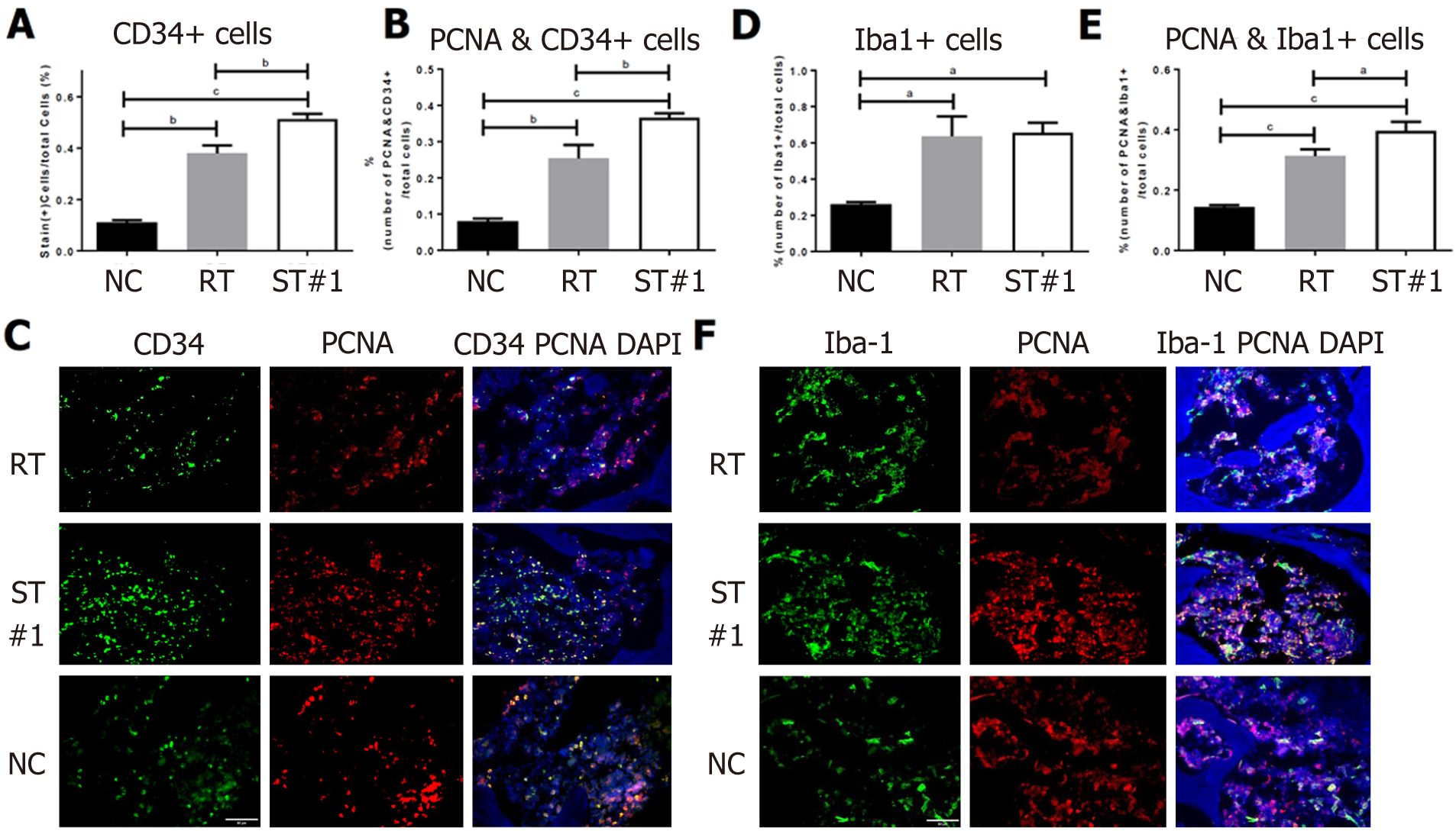
Figure 4 Immunohistochemical assessment of CD34, proliferating cell nuclear antigen, and ionized calcium-binding adaptor molecule-1 expression in bone marrow.
Three weeks following whole-body irradiation, mouse femurs were fixed, decalcified, and embedded in paraffin wax. The sections were deparaffinized, stained with antibodies, and imaged under 400 × magnification. A-C: The number of CD34+ cells in the stem cell-treated (ST)#1 group was higher than that in the irradiated (RT) group. The number of proliferating cell nuclear antigen (PCNA)+ cells in CD34+ hematopoietic stem cells was significantly higher in the ST#1 group than in the RT group; D-F: The number of ionized calcium-binding adaptor molecule-1 (Iba-1+) cells, a marker of monoblastic cells, was similar between the ST and RT groups; however, the number of cells expressing both PCNA and Iba-1 was significantly higher in the ST group. Magnification 400 ×; scale bar: 50 μm; aP < 0.001, bP < 0.001, and cP < 0.0001 vs controls. NC: Normal control; ST: Stem cell-treated; RT: Irradiated; CD: Cluster of differentiation; PCNA: Proliferating cell nuclear antigen; Iba-1: Ionized calcium-binding adaptor molecule-1; DAPI: 4′,6-diamidino-2-phenylindole.
- Citation: Kim MJ, Moon W, Heo J, Lim S, Lee SH, Jeong JY, Lee SJ. Optimization of adipose tissue-derived mesenchymal stromal cells transplantation for bone marrow repopulation following irradiation. World J Stem Cells 2022; 14(3): 245-263
- URL: https://www.wjgnet.com/1948-0210/full/v14/i3/245.htm
- DOI: https://dx.doi.org/10.4252/wjsc.v14.i3.245


