Copyright
©The Author(s) 2022.
World J Stem Cells. Feb 26, 2022; 14(2): 163-182
Published online Feb 26, 2022. doi: 10.4252/wjsc.v14.i2.163
Published online Feb 26, 2022. doi: 10.4252/wjsc.v14.i2.163
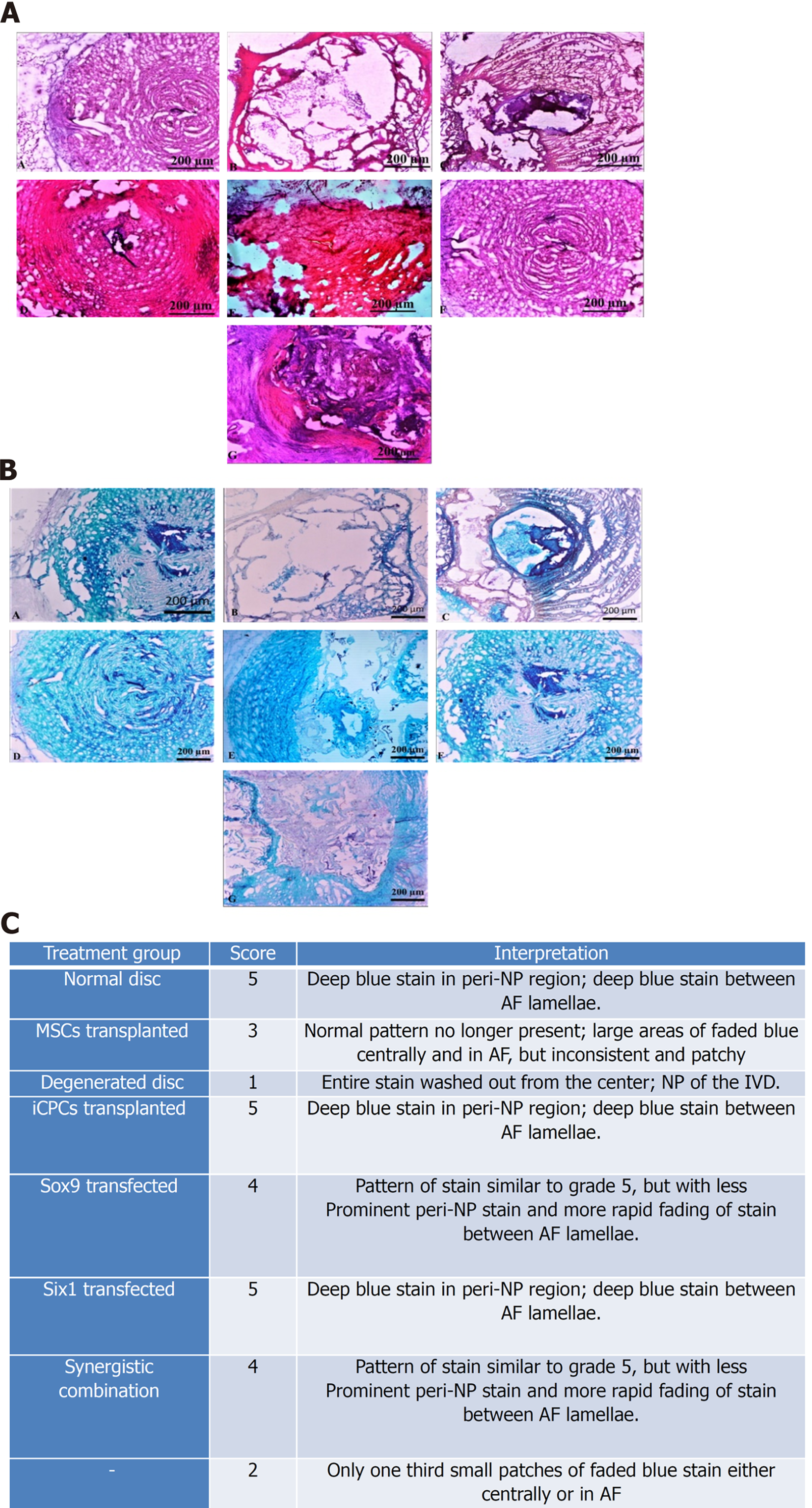
Figure 9 Histological examination of intervertebral disc.
A: (A-G) Bright field imaging of intervertebral discs (IVDs) showing nucleus pulposus (NP) content of the healthy (Co6/7), and degenerated disc (Co5/6) treated with normal mesenchymal stem cells (MSCs), transfected MSCs, and induced chondro-progenitor cells (iCPCs) (Co5/6) IVDs, in each group. Bright-field microscopic images of IVDs were captured from the cryosections stained by hematoxylin and eosin; normal healthy disc, degenerated disc, and degenerated disc transplanted with normal MSCs, Six-1 transfected MSCs, Sox-9 transfected MSCs, iCPCs, and Sox-9 + Six-1 transfected MSCs, respectively; B: (A-G) Bright-field microscopic images of IVDs were captured from the cryosections stained by Alcian blue displayed glycoproteins secreted by the transplanted cells compared to the degenerated disc; normal healthy disc, degenerated disc, and degenerated disc transplanted with normal MSCs, Six-1 transfected MSCs, Sox-9 transfected MSCs, iCPCs, and Sox-9 + Six-1 transfected MSCs, respectively; C: Histological grading with a score for regeneration. AF: Annular fibrosus.
- Citation: Khalid S, Ekram S, Salim A, Chaudhry GR, Khan I. Transcription regulators differentiate mesenchymal stem cells into chondroprogenitors, and their in vivo implantation regenerated the intervertebral disc degeneration. World J Stem Cells 2022; 14(2): 163-182
- URL: https://www.wjgnet.com/1948-0210/full/v14/i2/163.htm
- DOI: https://dx.doi.org/10.4252/wjsc.v14.i2.163


