Copyright
©The Author(s) 2021.
World J Stem Cells. Dec 26, 2021; 13(12): 1918-1927
Published online Dec 26, 2021. doi: 10.4252/wjsc.v13.i12.1918
Published online Dec 26, 2021. doi: 10.4252/wjsc.v13.i12.1918
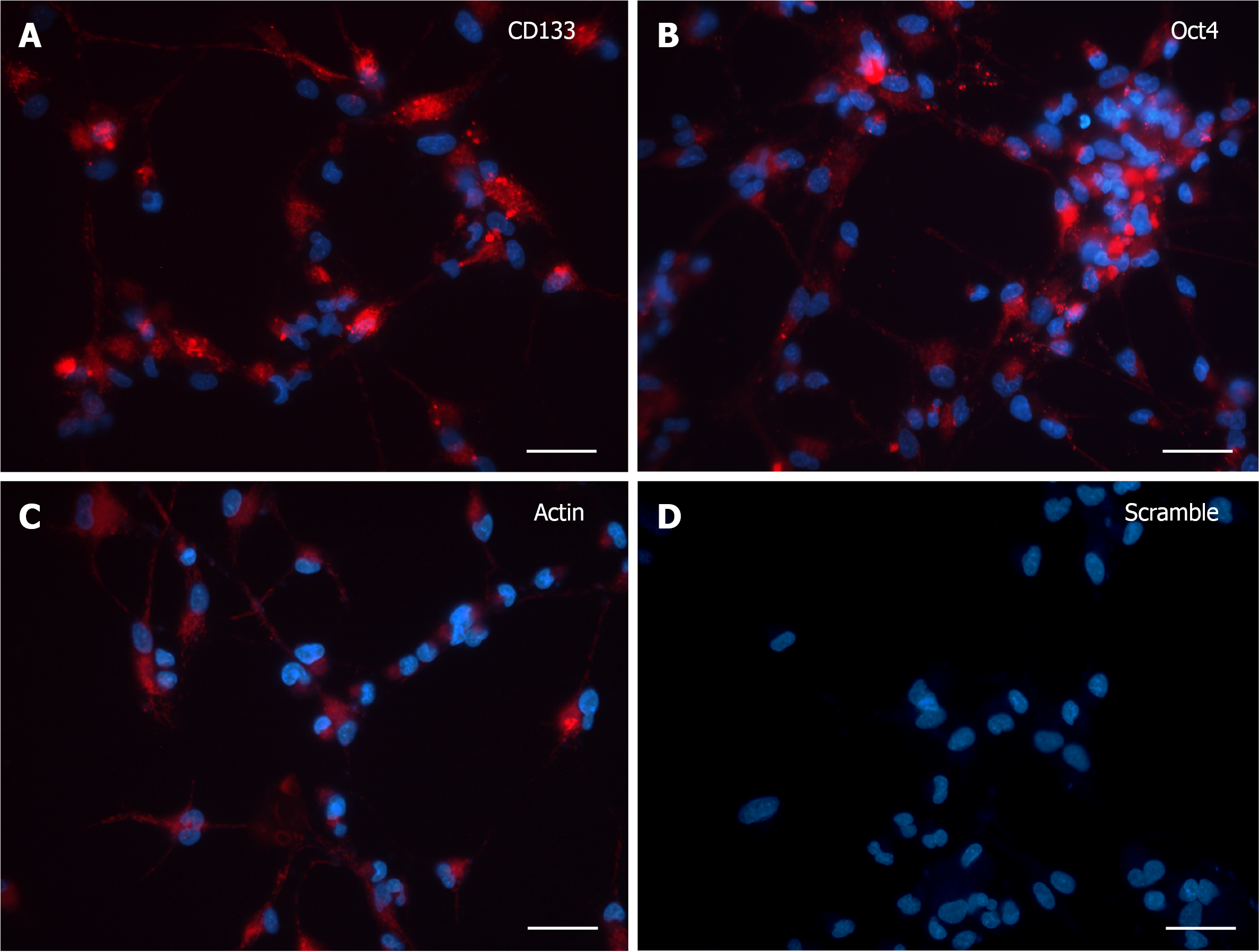
Figure 1 SmartFlareTM detection in 3 d in vitro neural stem cells.
A and B: The expression pattern for both SmartFlareTM CD133 and Oct4 probes showed a diffuse and spotted-like fluorescence from the perinuclear area to the peripheral cytological processes; C: SmartFlareTM probe for Actin showed a robust and overall localization of red fluorescence as indicative of positive mRNA expression; D: Scramble probe-treated cells were almost completely lacking in unspecific red background. Nuclei were stained by Hoechst dye. Scale bar: 50 μm.
- Citation: Diana A, Setzu MD, Kokaia Z, Nat R, Maxia C, Murtas D. SmartFlareTM is a reliable method for assessing mRNA expression in single neural stem cells. World J Stem Cells 2021; 13(12): 1918-1927
- URL: https://www.wjgnet.com/1948-0210/full/v13/i12/1918.htm
- DOI: https://dx.doi.org/10.4252/wjsc.v13.i12.1918


