Copyright
©The Author(s) 2021.
World J Stem Cells. Nov 26, 2021; 13(11): 1762-1782
Published online Nov 26, 2021. doi: 10.4252/wjsc.v13.i11.1762
Published online Nov 26, 2021. doi: 10.4252/wjsc.v13.i11.1762
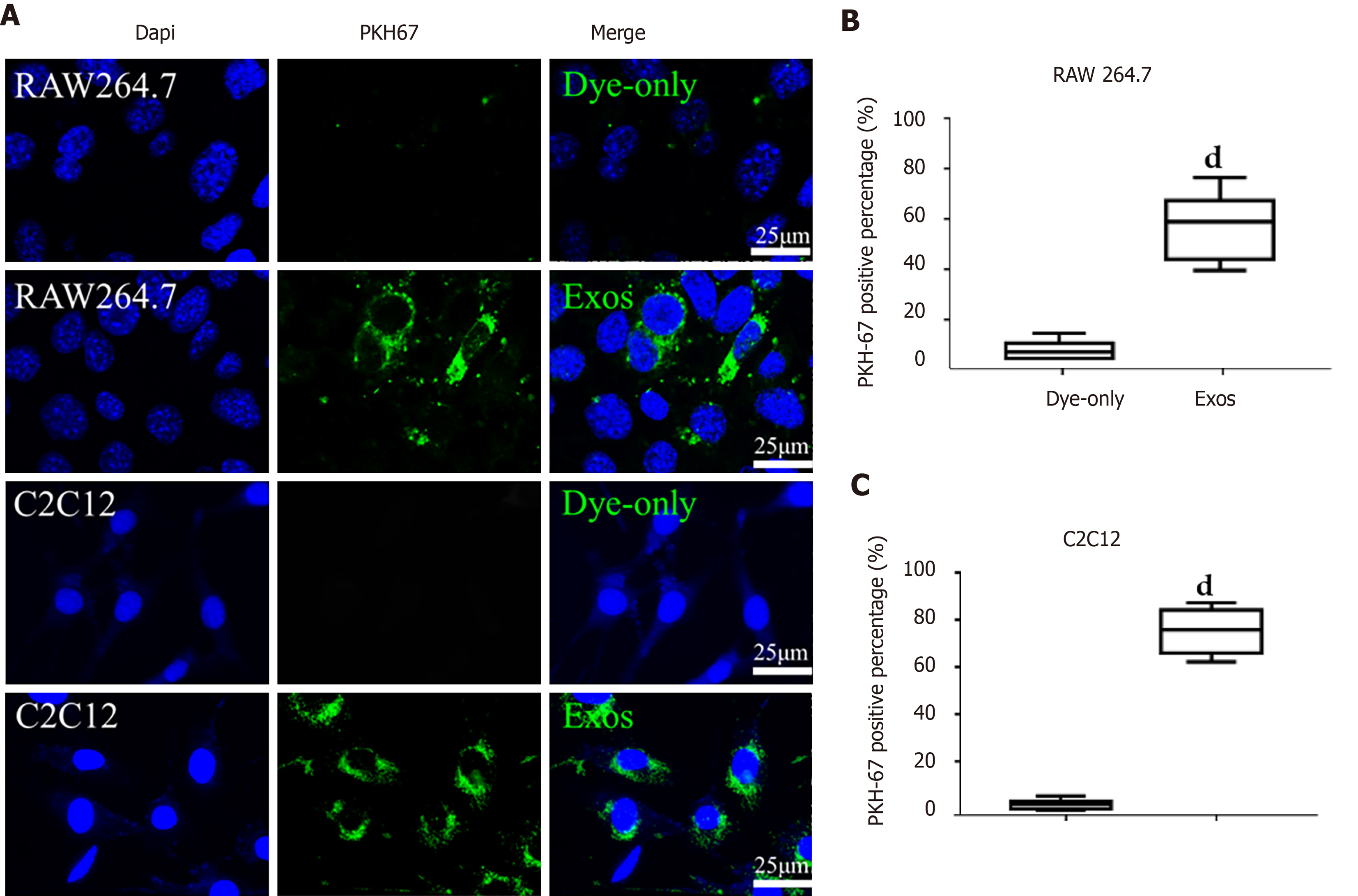
Figure 3 C2C12-Exos were taken up by C2C12 and RAW264.
7 cells in vitro. A: Representative images of the uptake of PKH67-labeled exosomes (green) by RAW264.7 cells or C2C12 cells (DAPI blue) and fluorescence uptake by negative control samples. Dye-only, only PKH67 incubated with RAW264.7 cells and C2C12 cells; scale bar = 25 μm; B and C: The percentage of PKH67 positive cells in RAW264.7 or C2C12 is presented. dP < 0.0001, n = 6.
- Citation: Luo ZW, Sun YY, Lin JR, Qi BJ, Chen JW. Exosomes derived from inflammatory myoblasts promote M1 polarization and break the balance of myoblast proliferation/differentiation. World J Stem Cells 2021; 13(11): 1762-1782
- URL: https://www.wjgnet.com/1948-0210/full/v13/i11/1762.htm
- DOI: https://dx.doi.org/10.4252/wjsc.v13.i11.1762


