Copyright
©The Author(s) 2021.
World J Stem Cells. Oct 26, 2021; 13(10): 1580-1594
Published online Oct 26, 2021. doi: 10.4252/wjsc.v13.i10.1580
Published online Oct 26, 2021. doi: 10.4252/wjsc.v13.i10.1580
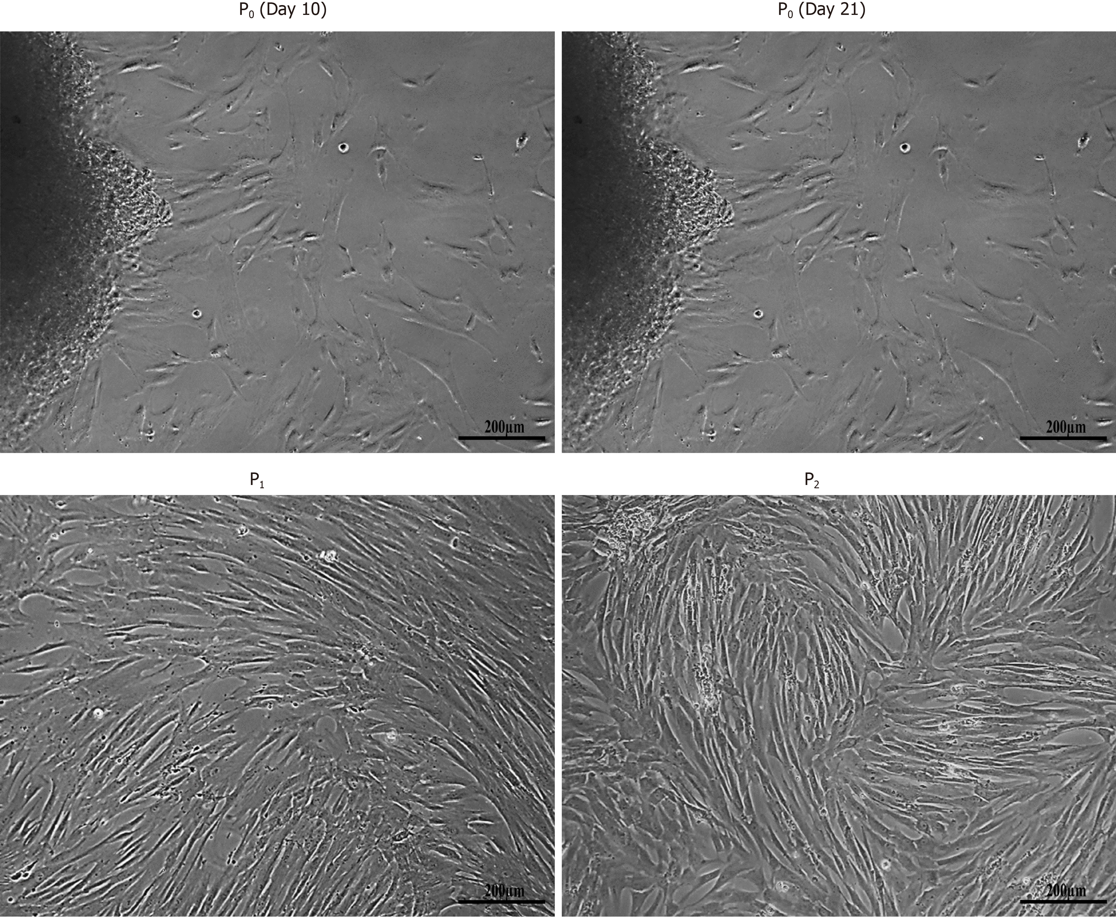
Figure 1 Isolation of human umbilical cord-mesenchymal stem cells.
Morphological observations of human umbilical cord-mesenchymal stem cells showing passage 0 (P0) cells at day 10 and day 21 with cells migrating towards the periphery of the adherent tissue, and passage 1 (P1) and passage 2 (P2) cells with homogeneous population showing fusiform- and fibroblast-like appearance after sub-culturing. All images were taken under phase contrast at 10× magnification.
- Citation: Fatima A, Malick TS, Khan I, Ishaque A, Salim A. Effect of glycyrrhizic acid and 18β-glycyrrhetinic acid on the differentiation of human umbilical cord-mesenchymal stem cells into hepatocytes. World J Stem Cells 2021; 13(10): 1580-1594
- URL: https://www.wjgnet.com/1948-0210/full/v13/i10/1580.htm
- DOI: https://dx.doi.org/10.4252/wjsc.v13.i10.1580


