Copyright
©The Author(s) 2021.
World J Stem Cells. Jan 26, 2021; 13(1): 78-90
Published online Jan 26, 2021. doi: 10.4252/wjsc.v13.i1.78
Published online Jan 26, 2021. doi: 10.4252/wjsc.v13.i1.78
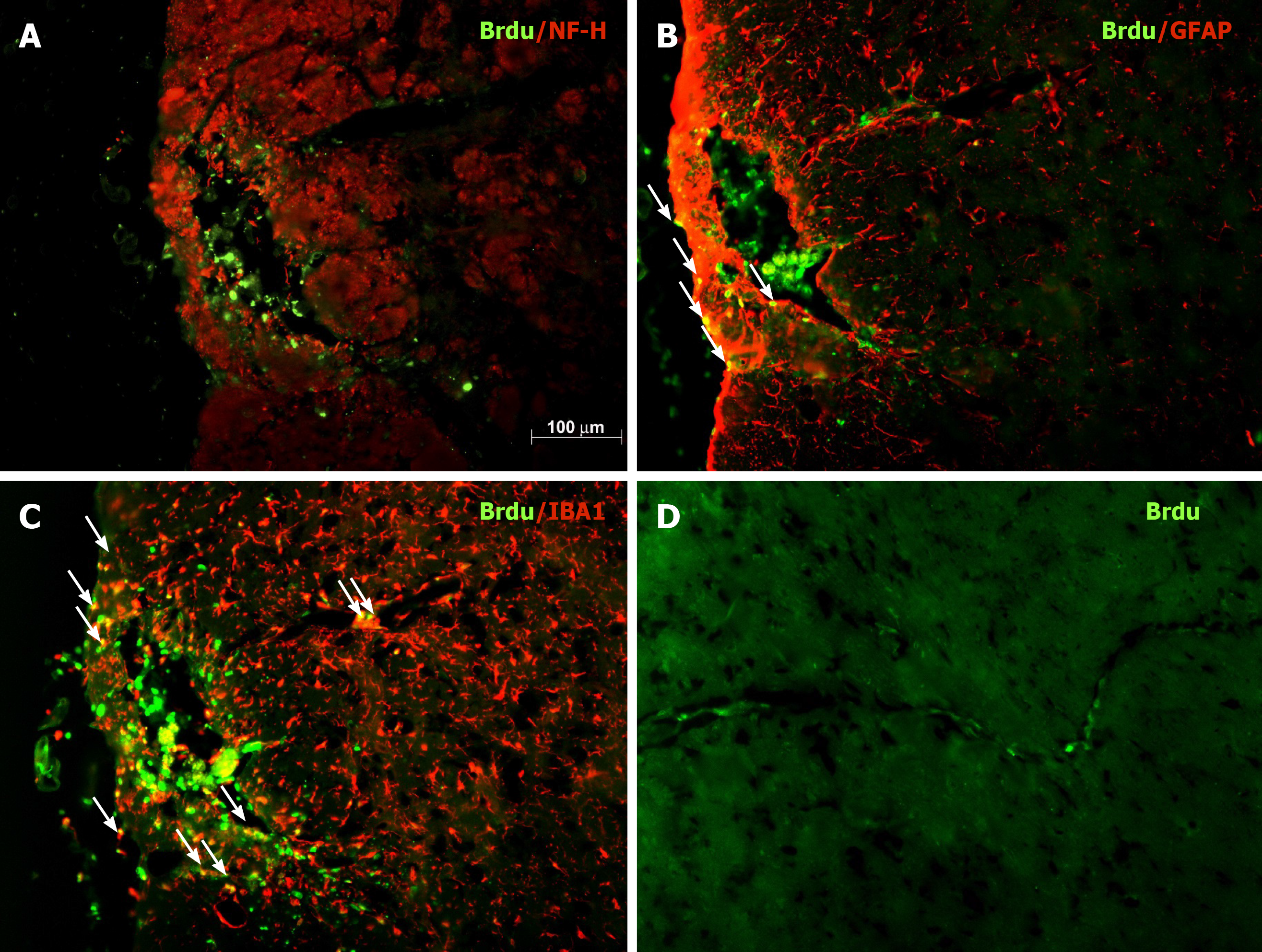
Figure 5 Phenotypic characterization of grafted bone marrow mesenchymal stem cells in the substantia nigra of hemiparkinsonian rats.
A-C: Representative micrographs of BrdU staining of coronal sections of the grafted substantia nigra. Photos of double staining (magnification, 200 ×) are shown. Green: BrdU immunoreactivity; red: NF-H immunoreactivity (neurons, panel A), GFAP immunoreactivity (astroglia, panel B), and IBA1 immunoreactivity (microglia, panel C); D: BrdU-immunoreactive BMSCs along the blood vessels near the graft site.
- Citation: Tsai MJ, Hung SC, Weng CF, Fan SF, Liou DY, Huang WC, Liu KD, Cheng H. Stem cell transplantation and/or adenoviral glial cell line-derived neurotrophic factor promote functional recovery in hemiparkinsonian rats. World J Stem Cells 2021; 13(1): 78-90
- URL: https://www.wjgnet.com/1948-0210/full/v13/i1/78.htm
- DOI: https://dx.doi.org/10.4252/wjsc.v13.i1.78


