Copyright
©The Author(s) 2021.
World J Stem Cells. Jan 26, 2021; 13(1): 115-127
Published online Jan 26, 2021. doi: 10.4252/wjsc.v13.i1.115
Published online Jan 26, 2021. doi: 10.4252/wjsc.v13.i1.115
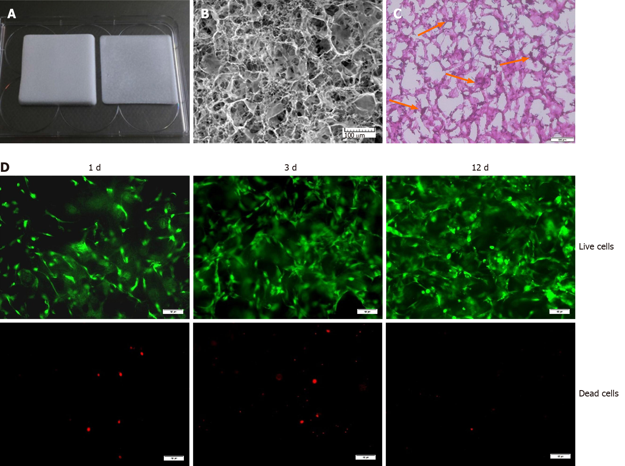
Figure 1 A 3D culture system based on a type I collagen sponge scaffold.
A: Type I collagen sponge scaffold; B: Surface topography of the collagen sponge scaffold observed by scanning electron microscopy. Bars = 100 μm; C: Hematoxylin and eosin staining of sectioned bone marrow mesenchymal stem cell-collagen sponge constructs. Orange arrows indicate the cells. Bars = 100 μm; D: Live/dead assay of the scaffolds during the cultivation process. Green indicates the live cells, and red indicates the dead cells. Bars = 100 μm.
- Citation: Zhang BY, Xu P, Luo Q, Song GB. Proliferation and tenogenic differentiation of bone marrow mesenchymal stem cells in a porous collagen sponge scaffold. World J Stem Cells 2021; 13(1): 115-127
- URL: https://www.wjgnet.com/1948-0210/full/v13/i1/115.htm
- DOI: https://dx.doi.org/10.4252/wjsc.v13.i1.115


