Copyright
©The Author(s) 2020.
World J Stem Cells. Aug 26, 2020; 12(8): 857-878
Published online Aug 26, 2020. doi: 10.4252/wjsc.v12.i8.857
Published online Aug 26, 2020. doi: 10.4252/wjsc.v12.i8.857
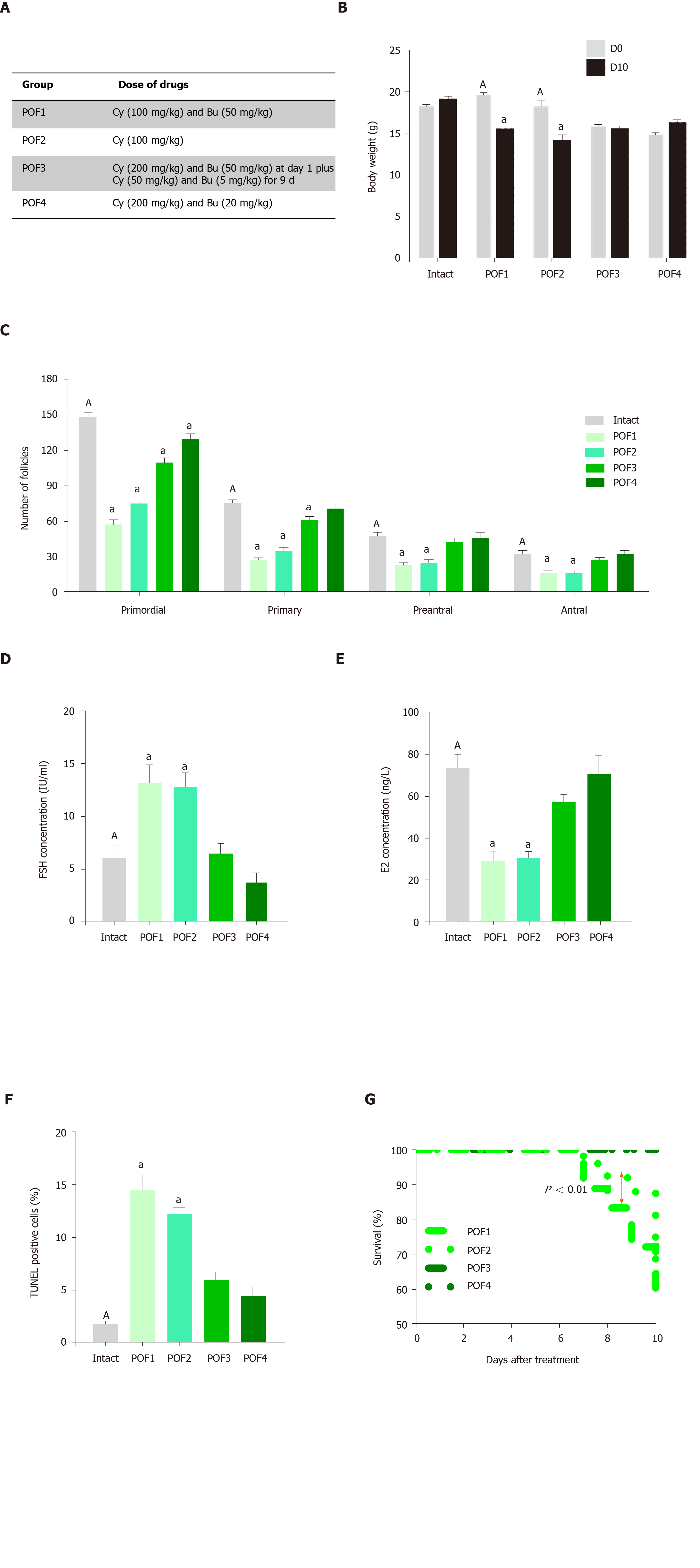
Figure 2 Establishment of a mouse model of premature ovarian failure.
A: Premature ovarian failure (POF) groups were treated with different dosages of cyclophosphamide and busulfan; B: Body weight changes in the intact and POF groups after 10 d showed that the POF1 and POF2 groups had significant decreases in body weights; C: Ovarian pathology of the intact and POF groups 10 d after injection of cyclophosphamide and busulfan . Follicle count revealed that there were fewer normal follicles in the POF groups than in the intact mice. The ovaries of the intact group contained large numbers of follicles at all developmental stages, whereas the atrophic ovaries of the POF groups had fewer follicles at each stage; D, E: Serum levels of follicle stimulating hormone and estradiol 10 d after injection of cyclophosphamide and busulfan. Serum levels of follicle stimulating hormone were significantly increased in the POF1 and POF2 groups compared to those of the intact group. Serum levels of estradiol were significantly decreased in the POF1 and POF2 groups compared with the intact group; F: Apoptosis rate in the ovary. Green fluorescence indicated the presence of apoptotic cells in the POF1, POF2, POF3 and POF4 groups; G: Survival rate in the POF groups after 10 d. The survival percent showed a significant decrease in the POF1 group compared with the POF2 group. All data are presented as mean ± standard error. Small letters (a) indicate the significance (P < 0.05) compared to groups labeled by similar capital letters (A); aP < 0.05 significance of experimental groups vs the intact group; n = 3-5. POF: Premature ovarian failure; Cy: Cyclophosphamide; Bu: Busulfan; FSH: Follicle stimulating hormone; E2: Estradiol.
- Citation: Bahrehbar K, Rezazadeh Valojerdi M, Esfandiari F, Fathi R, Hassani SN, Baharvand H. Human embryonic stem cell-derived mesenchymal stem cells improved premature ovarian failure. World J Stem Cells 2020; 12(8): 857-878
- URL: https://www.wjgnet.com/1948-0210/full/v12/i8/857.htm
- DOI: https://dx.doi.org/10.4252/wjsc.v12.i8.857


