Copyright
©The Author(s) 2020.
World J Stem Cells. Aug 26, 2020; 12(8): 857-878
Published online Aug 26, 2020. doi: 10.4252/wjsc.v12.i8.857
Published online Aug 26, 2020. doi: 10.4252/wjsc.v12.i8.857
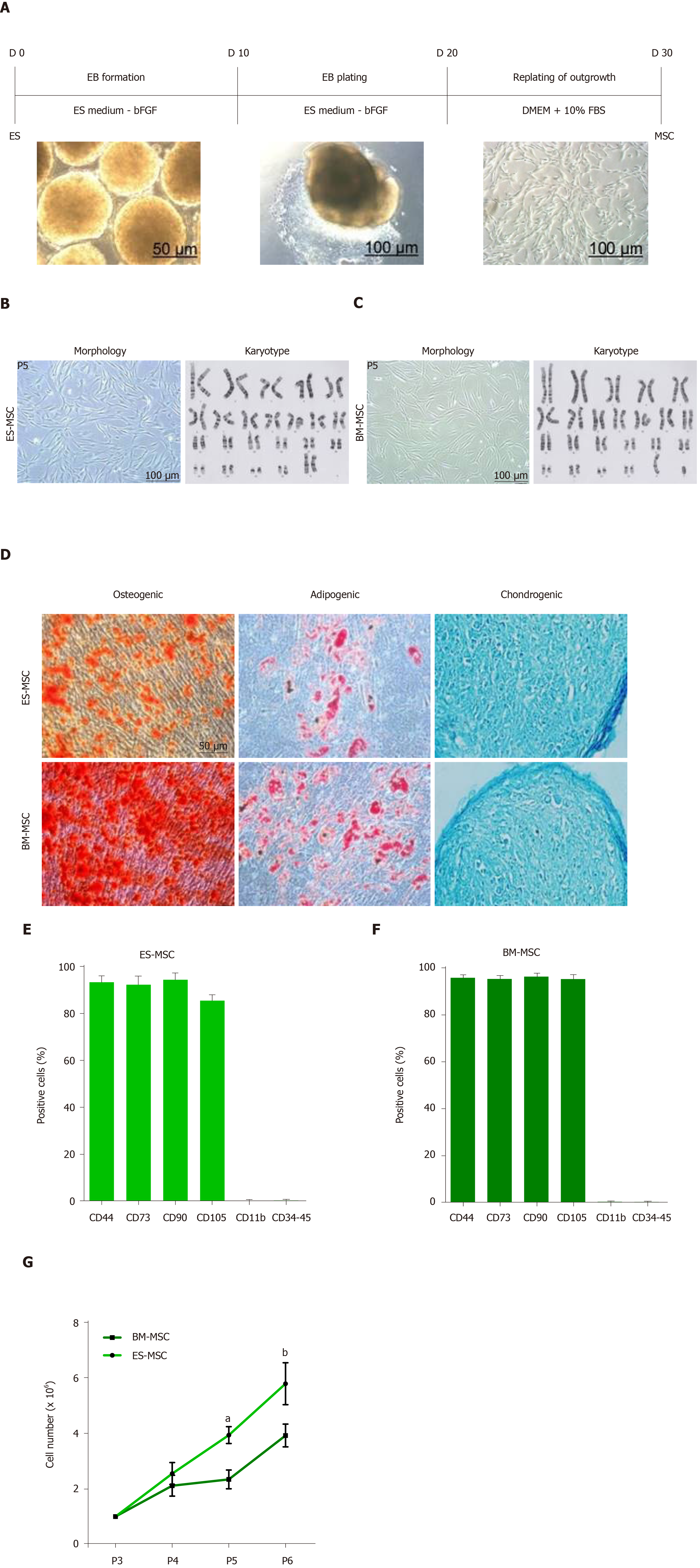
Figure 1 Derivation and identification of human embryonic stem cell-derived mesenchymal stem cells and bone marrow-derived mesenchymal stem cells.
A: Schematic presentation of the procedure used to derive human mesenchymal stem cells (MSCs) from embryonic stem (ES) cells. Colonies of ES cells were enzymatically detached and cultured for 10 d in suspension to form embryoid bodies, which were then plated onto gelatin-coated tissue culture plates. After 10 d, outgrowths of the cells that sprouted from embryoid bodies were mechanically isolated by a cell scraper and subsequently expanded in mesenchymal stem cell culture medium; B and C: Morphology and karyotype of ES-MSCs and BM-MSCs. Passage-5 ES-MSCs and BM-MSCs showed a fibroblastic morphology and normal karyotype; D: Alizarin red staining after 14 d of culture in osteogenic medium indicated the osteogenic differentiation potential of ES-MSCs and BM-MSCs (P4). Oil red staining after 21 d of culture in adipogenic medium showed the adipogenic differentiation potential of ES-MSCs and BM-MSCs (P4). Alcian blue staining after 21 d of culture in chondrogenic medium showed chondrogenic differentiation potential of ES-MSCs and BM-MSCs; E, F: Flow cytometric analysis indicated that cultured ES-MSCs and BM-MSCs expressed CD44, CD90, CD73 and endoglin (CD105), but not hematopoietic lineage markers CD11b, CD34 and protein tyrosine phosphatase receptor type C (CD45); G: ES-MSCs proliferated more rapidly than BM-MSCs. Results are expressed as mean ± standard error,
- Citation: Bahrehbar K, Rezazadeh Valojerdi M, Esfandiari F, Fathi R, Hassani SN, Baharvand H. Human embryonic stem cell-derived mesenchymal stem cells improved premature ovarian failure. World J Stem Cells 2020; 12(8): 857-878
- URL: https://www.wjgnet.com/1948-0210/full/v12/i8/857.htm
- DOI: https://dx.doi.org/10.4252/wjsc.v12.i8.857


