Copyright
©The Author(s) 2020.
World J Stem Cells. Dec 26, 2020; 12(12): 1652-1666
Published online Dec 26, 2020. doi: 10.4252/wjsc.v12.i12.1652
Published online Dec 26, 2020. doi: 10.4252/wjsc.v12.i12.1652
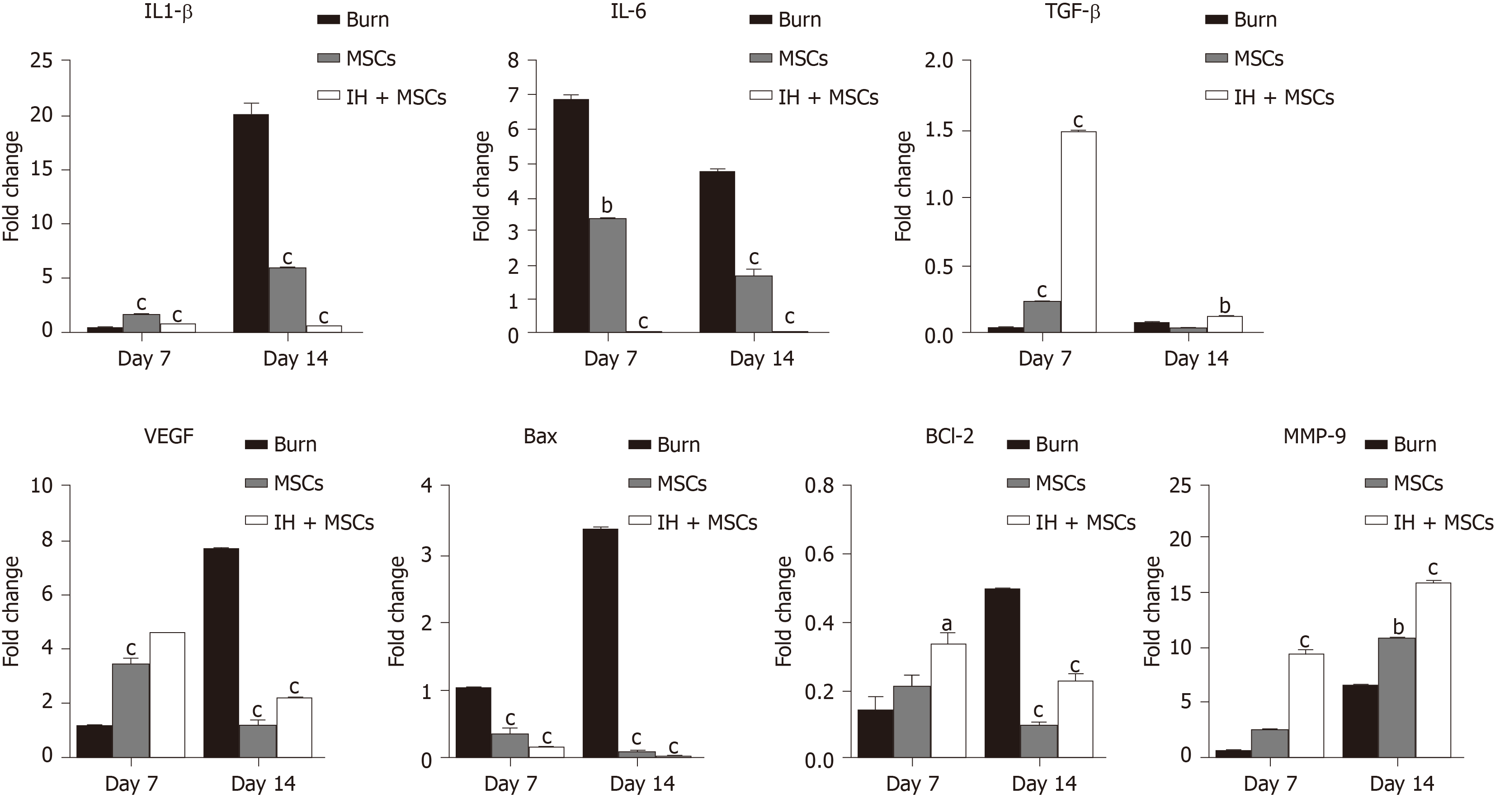
Figure 6 Gene expression analysis of burn wounds.
Bar diagrams corresponding to gene expression analysis of interleukin (IL)-1β, IL-6, transforming growth factor β (TGF-β), vascular endothelial growth factor (VEGF), Bcl-2 associated X (Bax), Bcl-2 and matrix metallopeptidase 9 (MMP-9) are shown in the burn wound control (at days 7 and 14) and experimental groups treated with normal untreated mesenchymal stem cells (MSCs) and preconditioned MSCs (IH + MSCs) at days 7 and 14 following 72 h of burn injury. The pattern of gene expression was time dependent; statistically significant downregulation of IL-1β (at day 14), IL-6 and Bax (days 7 and 14), and significant upregulation of TGF-β and MMP-9 (days 7 and 14), VEGF and Bcl-2 (day 7) in case of IH + MSCs as compared to burn control. Same pattern was observed in the case of normal MSCs for IL-1β, IL-6, Bax VEGF and Bcl-2 genes, while for TGF-β (day 14) and MMP-9 (day 7), the expression levels were non-significant. For statistical analysis, One way ANOVA was performed, followed by the Bonferroni post hoc test. Data are presented as the mean ± SE (n = 3). aP < 0.05, bP < 0.01 and cP < 0.001. IL: Interleukin; TGF-β: Transforming growth factor β; VEGF: Vascular endothelial growth factor; MMP-9: Matrix metalloproteinase-9.
- Citation: Aslam S, Khan I, Jameel F, Zaidi MB, Salim A. Umbilical cord-derived mesenchymal stem cells preconditioned with isorhamnetin: potential therapy for burn wounds. World J Stem Cells 2020; 12(12): 1652-1666
- URL: https://www.wjgnet.com/1948-0210/full/v12/i12/1652.htm
- DOI: https://dx.doi.org/10.4252/wjsc.v12.i12.1652


