Copyright
©The Author(s) 2020.
World J Stem Cells. Dec 26, 2020; 12(12): 1652-1666
Published online Dec 26, 2020. doi: 10.4252/wjsc.v12.i12.1652
Published online Dec 26, 2020. doi: 10.4252/wjsc.v12.i12.1652
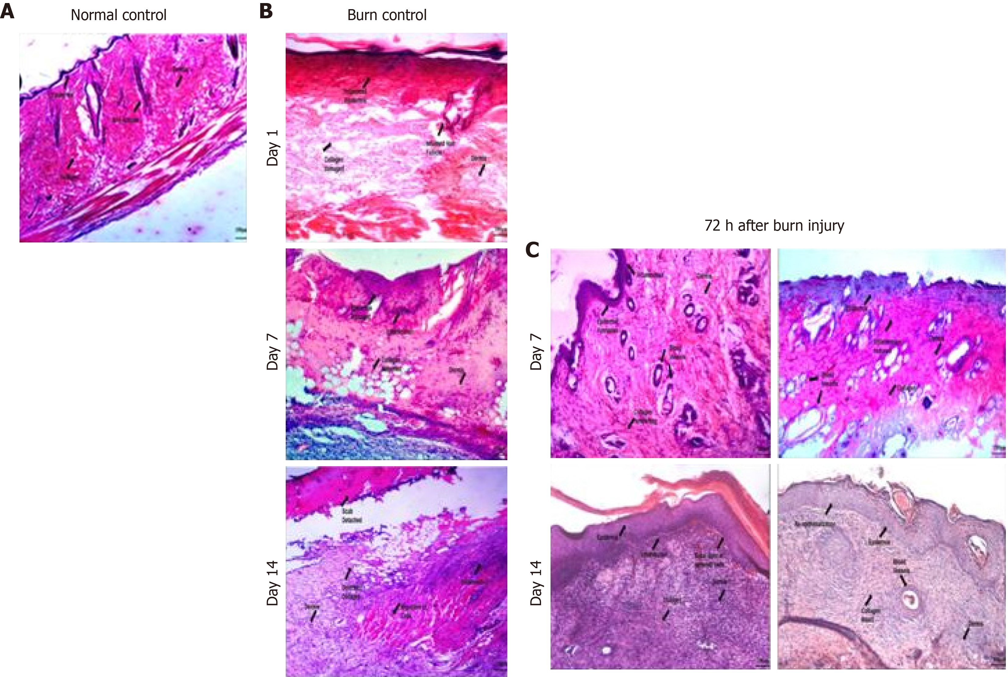
Figure 5 Histological analysis of normal and burn wound tissues.
Phase contrast images (20 ×) of histological sections stained with hematoxylin-eosin stain showing normal control (skin with no wound) with normal skin structure composed of the epidermis, dermis and hypodermis along with the muscles, burn wound control at days 1, 7 and 14, and burn wound tissues transplanted with normal mesenchymal stem cells (MSCs) and preconditioned MSCs (IH + MSCs) at days 7 and 14 following 72 h of burn injury. The burn wound at day 1 shows inflamed epidermis and hair follicles, completely distorted dermis and collagen matrix; at day 7, disorganized tissue framework with no distinct layers of skin and the presence of loose connective tissues, while hair follicles show better morphology and reduced inflammation; and at day 14, slight detachment of the scab with deformed collagen and cellular infiltration, loose connective tissue with disorganized dermis and absence of epidermis. Normal MSCs treated burn wound at day 7 shows neovascularization and inflamed epidermis along with collagen deposition, while hair follicles show inflammatory cell infiltration in the periphery with better morphology; and at day 14, epidermis formation with cellular migration and collagen deposition, constructing the dermis. IH + MSCs treated burn wound at day 7 shows increased angiogenesis and reduced inflammation in the epidermis, while hair follicles also show similar morphology to that in normal skin with reduced inflammation; and at day 14, complete re-epithelialization with intact collagen and vascularization, and remodeling of the tissue framework with a distinct layer of epidermis and dermis with no signs of scar. A: Normal control; B: Burn control; C: Seventy two hours (72 h) after burn injury.
- Citation: Aslam S, Khan I, Jameel F, Zaidi MB, Salim A. Umbilical cord-derived mesenchymal stem cells preconditioned with isorhamnetin: potential therapy for burn wounds. World J Stem Cells 2020; 12(12): 1652-1666
- URL: https://www.wjgnet.com/1948-0210/full/v12/i12/1652.htm
- DOI: https://dx.doi.org/10.4252/wjsc.v12.i12.1652


