Copyright
©The Author(s) 2020.
World J Stem Cells. Dec 26, 2020; 12(12): 1652-1666
Published online Dec 26, 2020. doi: 10.4252/wjsc.v12.i12.1652
Published online Dec 26, 2020. doi: 10.4252/wjsc.v12.i12.1652
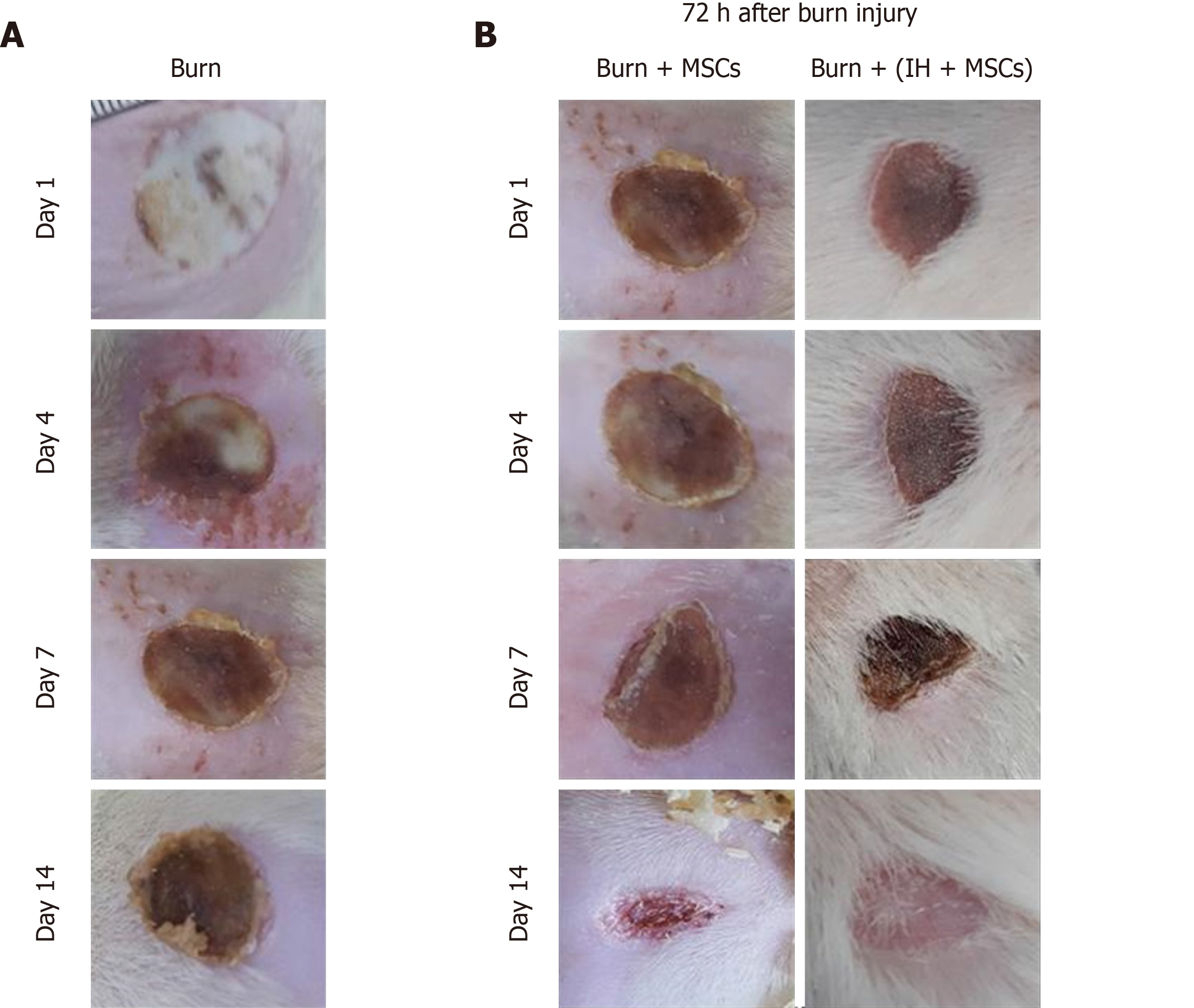
Figure 4 Macroscopic examination of burn wounds.
A: Images of burn wound tissue visualized macroscopically at days 1, 4, 7 and 14. Scab formation was observed at day 4 and slight detachment of the scab at day 14; B: Burn wound tissues transplanted with normal mesenchymal stem cells (MSCs) and preconditioned MSCs (IH + MSCs) were visualized 72 h after burn injury i.e. at day 1, 4, 7 and 14 following transplantation. At day 4, wound progression and initiation of scab formation were observed in all transplantation groups. At day 7, a thick scab with slight detachment was observed which completely covered the burn site and no sign of infection in the group transplanted with IH + MSCs. At day 14, complete detachment of the scab with slight granulation tissue was observed in the group transplanted with normal MSCs, while complete scab detachment and re-epithelialization were observed along with hair growth in the case of IH + MSCs. MSCs: Mesenchymal stem cells; IH + MSCs: Preconditioned mesenchymal stem cells.
- Citation: Aslam S, Khan I, Jameel F, Zaidi MB, Salim A. Umbilical cord-derived mesenchymal stem cells preconditioned with isorhamnetin: potential therapy for burn wounds. World J Stem Cells 2020; 12(12): 1652-1666
- URL: https://www.wjgnet.com/1948-0210/full/v12/i12/1652.htm
- DOI: https://dx.doi.org/10.4252/wjsc.v12.i12.1652


