
















©The Author(s) 2020.
World J Stem Cells. Dec 26, 2020; 12(12): 1652-1666
Published online Dec 26, 2020. doi: 10.4252/wjsc.v12.i12.1652
Published online Dec 26, 2020. doi: 10.4252/wjsc.v12.i12.1652
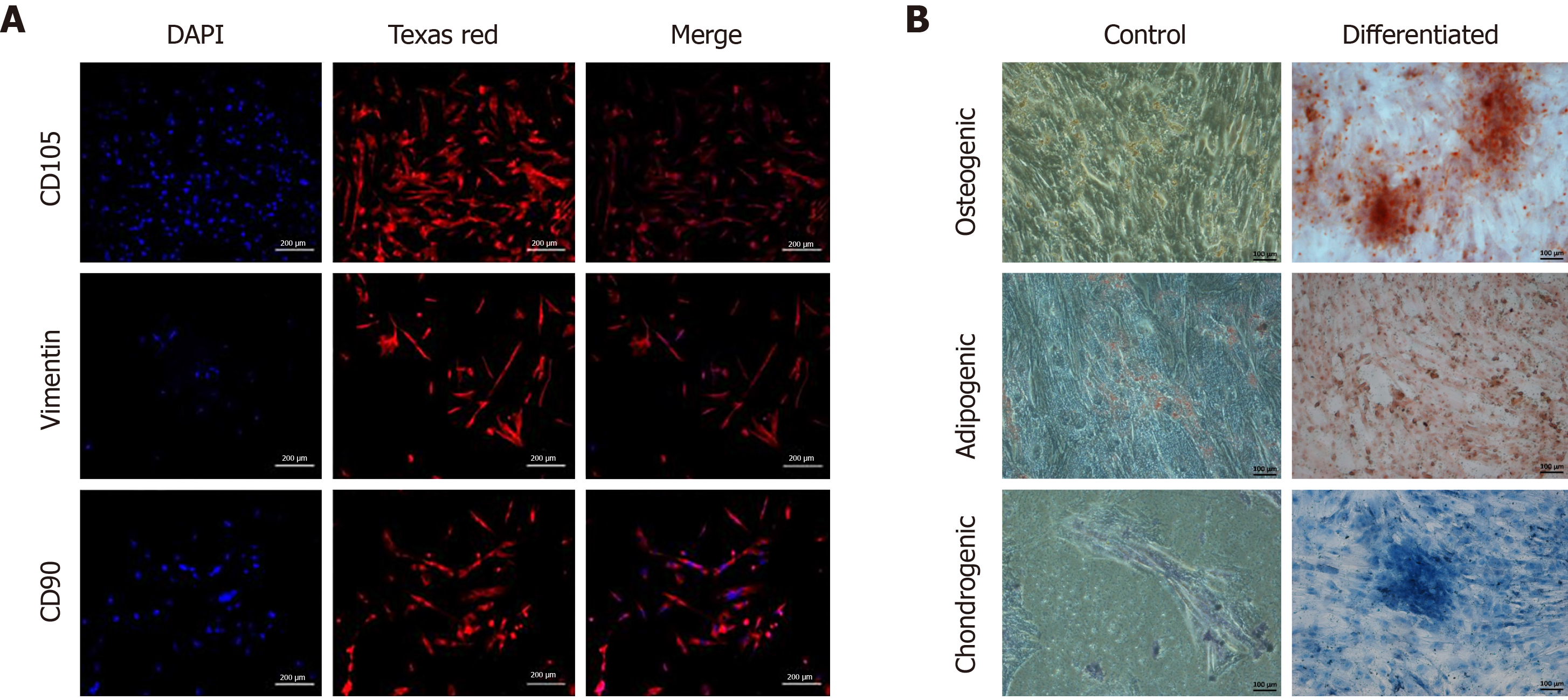
Figure 2 Characterization of mesenchymal stem cells.
A: Fluorescent images (10 ×) of isolated mesenchymal stem cells (MSCs) characterized by immunocytochemistry showing positive expression of cell surface markers CD105, vimentin and CD90. Alexa Fluor 546 goat anti-mouse secondary antibody was used for detection. Nuclei were stained with DAPI; B: Phase contrast images (10 ×) of MSCs stained with Alizarin Red stain showing mineral deposits, Oil Red O stain showing oil droplets, and Alcian Blue stain showing proteoglycans, confirming trilineage (osteogenic, adipogenic and chondrogenic) differentiation of MSCs grown in specific differentiation medium, as compared to the control.
- Citation: Aslam S, Khan I, Jameel F, Zaidi MB, Salim A. Umbilical cord-derived mesenchymal stem cells preconditioned with isorhamnetin: potential therapy for burn wounds. World J Stem Cells 2020; 12(12): 1652-1666
- URL: https://www.wjgnet.com/1948-0210/full/v12/i12/1652.htm
- DOI: https://dx.doi.org/10.4252/wjsc.v12.i12.1652