Copyright
©The Author(s) 2020.
World J Stem Cells. Oct 26, 2020; 12(10): 1133-1151
Published online Oct 26, 2020. doi: 10.4252/wjsc.v12.i10.1133
Published online Oct 26, 2020. doi: 10.4252/wjsc.v12.i10.1133
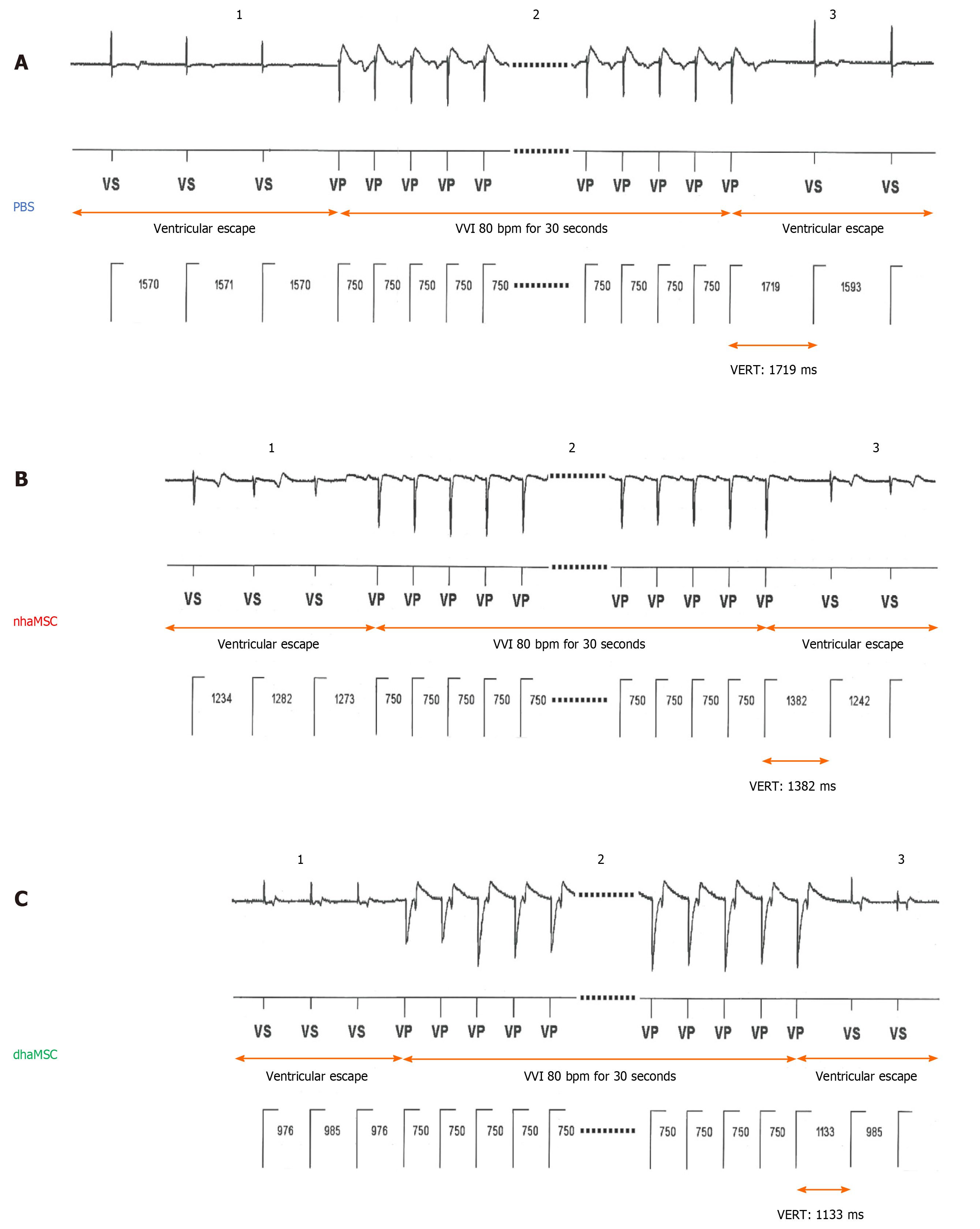
Figure 6 Ventricular escape rhythm recovery time after atrioventricular node ablation.
A: Phosphate-buffered saline (PBS) group; B: Native human mesenchymal stem cells derived from adipose tissue (nhaMSC) group; C: Differentiated human mesenchymal stem cells derived from adipose tissue (dhaMSC) group. 1: Ventricular escape rhythm before overstimulation by electronic pacemaker. 2: Overstimulation with VVI 80 bpm for 30 s by electronic pacemaker. 3: Ventricular escape rhythm after overstimulation by electronic pacemaker. Depicted ventricular escape rhythm recovery time values are exemplary for one pig of each group (dhaMSC, nhaMSC or PBS), average values of n = 6 pigs are included in the text. Electrocardiograms recorded at a speed of 25 mm/s. PBS: Phosphate-buffered saline; nhaMSC: Native human mesenchymal stem cells derived from adipose tissue; dhaMSC: Differentiated human mesenchymal stem cells derived from adipose tissue; VERT: Ventricular escape rhythm recovery time.
- Citation: Darche FF, Rivinius R, Rahm AK, Köllensperger E, Leimer U, Germann G, Reiss M, Koenen M, Katus HA, Thomas D, Schweizer PA. In vivo cardiac pacemaker function of differentiated human mesenchymal stem cells from adipose tissue transplanted into porcine hearts. World J Stem Cells 2020; 12(10): 1133-1151
- URL: https://www.wjgnet.com/1948-0210/full/v12/i10/1133.htm
- DOI: https://dx.doi.org/10.4252/wjsc.v12.i10.1133


