Copyright
©The Author(s) 2020.
World J Stem Cells. Jan 26, 2020; 12(1): 35-54
Published online Jan 26, 2020. doi: 10.4252/wjsc.v12.i1.35
Published online Jan 26, 2020. doi: 10.4252/wjsc.v12.i1.35
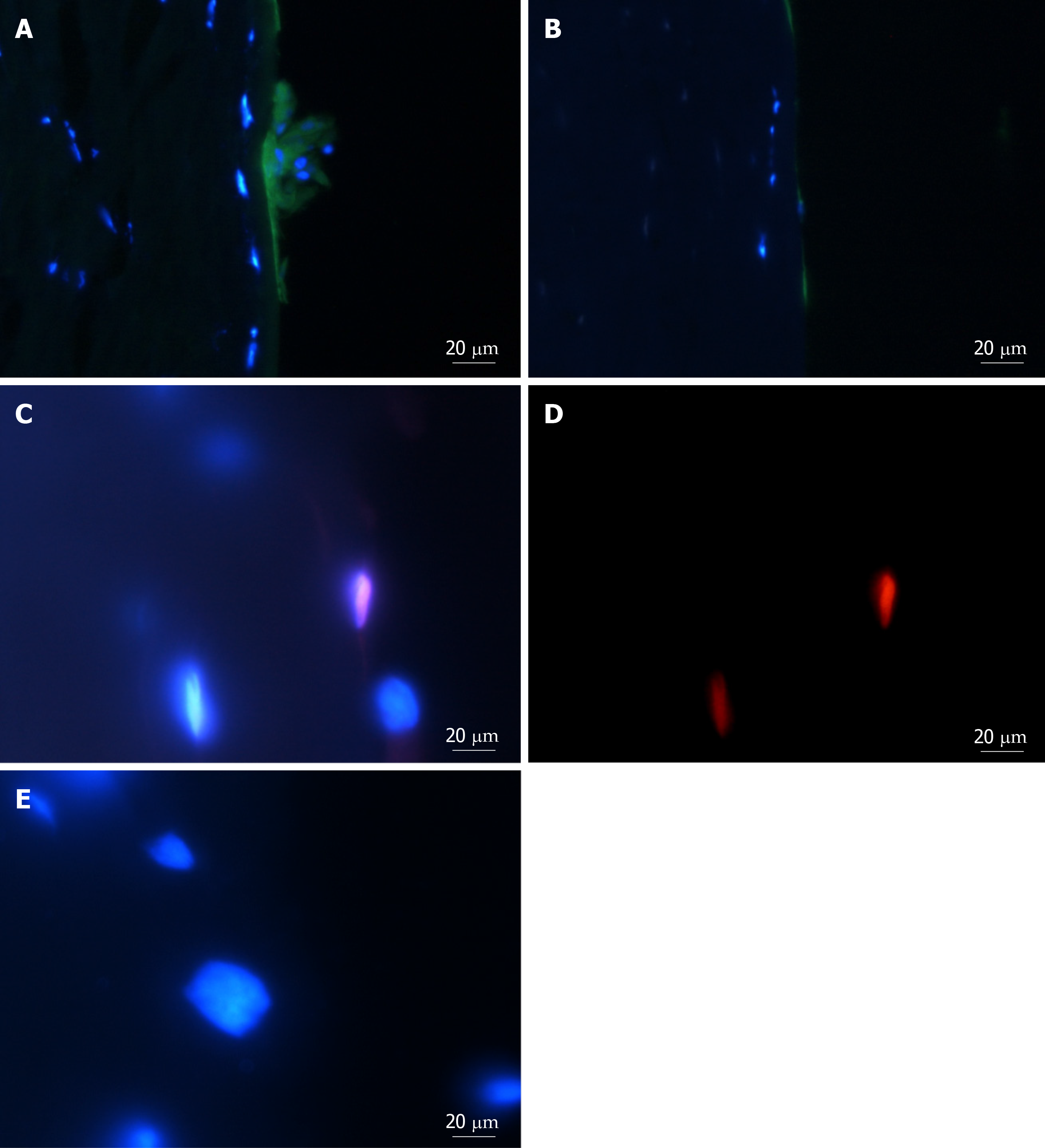
Figure 6 Putative stem-cell marker tumour protein p63, transcript variant 1, N-terminal isoform and cell-proliferation marker 5-ethynyl-2'-deoxyuridine in cells within sphere-implanted-full-thickness tissue at day 10.
A: Representative images of cross sections with spheres and cells derived from spheres implanted into full-thickness keratoconic and non-keratoconic central corneal buttons at day 10 show positive staining within the sphere for the putative stem cell marker tumour protein p63, transcript variant 1, N-terminal isoform (∆Np63α) (green); B: Cells stained without primary antibody (secondary antibody only control) show low green fluorescence. Cell nuclei are stained with DAPI (blue); C, D: Some, but not all cells show evidence of cell division (detected with ClickiT 5-ethynyl-2'-deoxyuridine (EdU) (red), a cell proliferation marker), as shown by pink cell nuclei in superimposed images of DAPI and EdU staining (C) and its equivalent image with red signal only (D); E: A negative control (EdU incubation without processing with its reaction solution) imaged at the same signal intensity as D is shown superimposed on DAPI for comparison. Scale bar = 20 µm.
- Citation: Wadhwa H, Ismail S, McGhee JJ, Van der Werf B, Sherwin T. Sphere-forming corneal cells repopulate dystrophic keratoconic stroma: Implications for potential therapy. World J Stem Cells 2020; 12(1): 35-54
- URL: https://www.wjgnet.com/1948-0210/full/v12/i1/35.htm
- DOI: https://dx.doi.org/10.4252/wjsc.v12.i1.35


