Copyright
©The Author(s) 2020.
World J Stem Cells. Jan 26, 2020; 12(1): 35-54
Published online Jan 26, 2020. doi: 10.4252/wjsc.v12.i1.35
Published online Jan 26, 2020. doi: 10.4252/wjsc.v12.i1.35
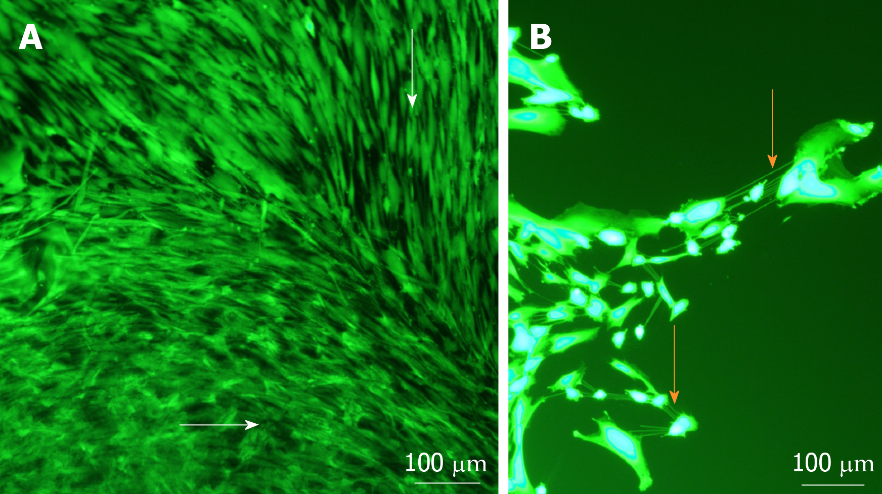
Figure 3 Cell morphology and migration patterns on the surface of sphere-seeded en face keratoconic tissue sections.
A: Representative images of migrated cells over the surface of keratoconic tissue sections show cells from the centre of the sphere aligned radially while cells close to the tissue edge align parallel with the tissue edge (white arrows show cell orientations); B: When cells at one of the migrating edges are magnified and overexposed, fine cell projections can be seen connecting cells to neighbouring ones (orange arrows). Scale bar = 100 µm.
- Citation: Wadhwa H, Ismail S, McGhee JJ, Van der Werf B, Sherwin T. Sphere-forming corneal cells repopulate dystrophic keratoconic stroma: Implications for potential therapy. World J Stem Cells 2020; 12(1): 35-54
- URL: https://www.wjgnet.com/1948-0210/full/v12/i1/35.htm
- DOI: https://dx.doi.org/10.4252/wjsc.v12.i1.35


