Copyright
©The Author(s) 2019.
World J Stem Cells. Feb 26, 2019; 11(2): 124-146
Published online Feb 26, 2019. doi: 10.4252/wjsc.v11.i2.124
Published online Feb 26, 2019. doi: 10.4252/wjsc.v11.i2.124
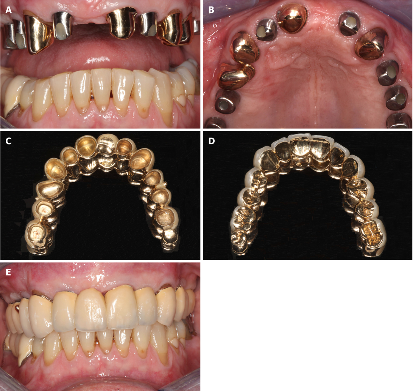
Figure 8 Documentation of clinical outcome.
A, B: Intraoral ventral (A) and occlusal (B) views on the healing abutments in regions 12, 14, 15, 16, 22, 25, and 26 after the fourth treatment (1 year after the placement of implants and 20 mo after guided bone regeneration maxillary sinus augmentation). Note that the teeth # 13, 21, 23, and 24 are crowned; C, D: External (C) and internal (D) view of the prosthetic telescopic bridge; E: Intraoral view of the final prosthetic reconstruction.
- Citation: Solakoglu Ö, Götz W, Kiessling MC, Alt C, Schmitz C, Alt EU. Improved guided bone regeneration by combined application of unmodified, fresh autologous adipose derived regenerative cells and plasma rich in growth factors: A first-in-human case report and literature review. World J Stem Cells 2019; 11(2): 124-146
- URL: https://www.wjgnet.com/1948-0210/full/v11/i2/124.htm
- DOI: https://dx.doi.org/10.4252/wjsc.v11.i2.124


