Copyright
©The Author(s) 2019.
World J Stem Cells. Feb 26, 2019; 11(2): 124-146
Published online Feb 26, 2019. doi: 10.4252/wjsc.v11.i2.124
Published online Feb 26, 2019. doi: 10.4252/wjsc.v11.i2.124
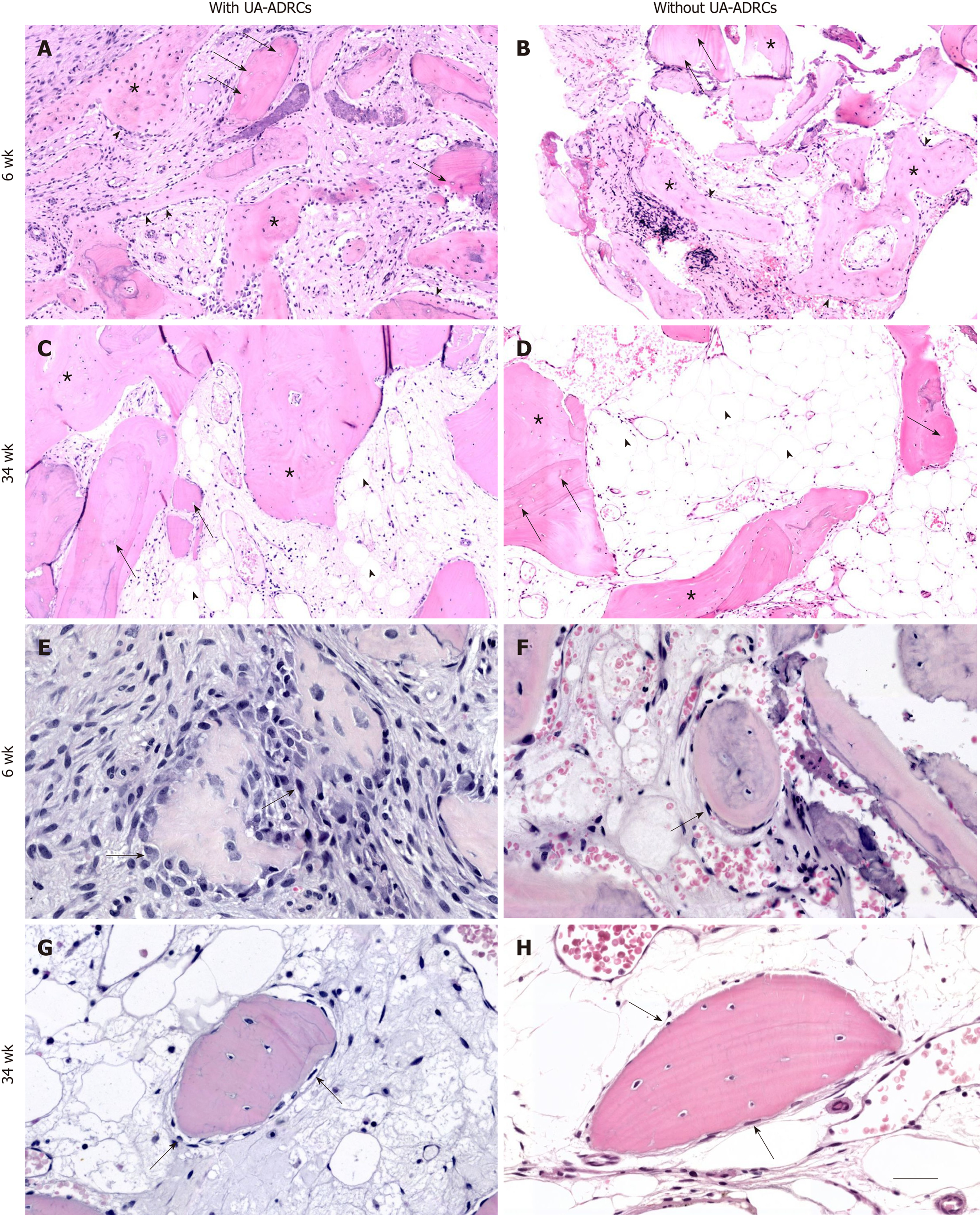
Figure 5 Histological findings.
Representative photomicrographs of 3 µm thick paraffin sections stained with hematoxylin and eosin of biopsies that were collected at 6 wk (A, B, E, F) or 34 wk (C, D, G, H) after guided bone regeneration maxillary sinus augmentation with the application of UA-ADRCs (A, C, E, G) or without UA-ADRCs (B, D, F, H). In A-D the asterisks indicate newly formed bone and the arrows indicate empty osteocyte lacunae in allogeneic fragments. Furthermore, in A and B the arrowheads point to osteoblasts with underlying osteoid, while in C and D the arrowheads indicate adipocytes. In E-H the arrows indicate cells on the surface of newly formed bone. The scale bar in H represents 150 µm in A-D and 75 µm in E-H. UA-ADRCs: Unmodified autologous adipose-derived regenerative cells.
- Citation: Solakoglu Ö, Götz W, Kiessling MC, Alt C, Schmitz C, Alt EU. Improved guided bone regeneration by combined application of unmodified, fresh autologous adipose derived regenerative cells and plasma rich in growth factors: A first-in-human case report and literature review. World J Stem Cells 2019; 11(2): 124-146
- URL: https://www.wjgnet.com/1948-0210/full/v11/i2/124.htm
- DOI: https://dx.doi.org/10.4252/wjsc.v11.i2.124


