Copyright
©The Author(s) 2019.
World J Stem Cells. Feb 26, 2019; 11(2): 124-146
Published online Feb 26, 2019. doi: 10.4252/wjsc.v11.i2.124
Published online Feb 26, 2019. doi: 10.4252/wjsc.v11.i2.124
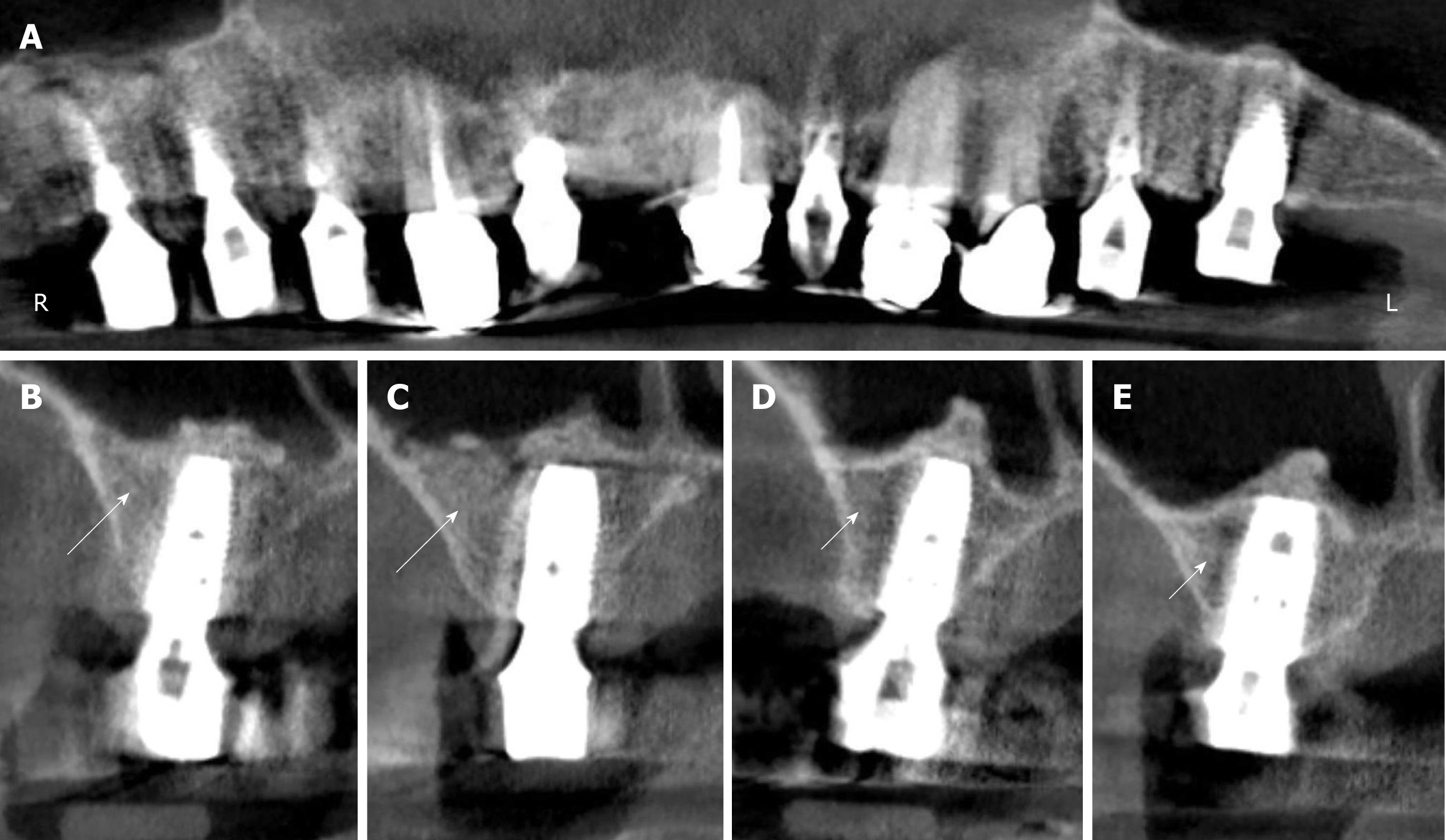
Figure 4 Digital volume tomography radiographs taken after the fourth surgery (1 year after the placement of implants and 20 mo after guided bone regeneration-maxillary sinus augmentation).
A: Panoramic view; B-E: Detailed view on selected regions 15 (B), 16 (C), 25 (D), and 26 (E). With the application of unmodified autologous adipose-derived regenerative cells (UA-ADRCs) the bone around the implants in regions 15 and 16 appeared larger in area and denser (arrows in B, C) than without the application of UA-ADRCs in regions 25 and 26 (arrowheads in D, E).
- Citation: Solakoglu Ö, Götz W, Kiessling MC, Alt C, Schmitz C, Alt EU. Improved guided bone regeneration by combined application of unmodified, fresh autologous adipose derived regenerative cells and plasma rich in growth factors: A first-in-human case report and literature review. World J Stem Cells 2019; 11(2): 124-146
- URL: https://www.wjgnet.com/1948-0210/full/v11/i2/124.htm
- DOI: https://dx.doi.org/10.4252/wjsc.v11.i2.124


