Copyright
©The Author(s) 2018.
World J Stem Cells. Nov 26, 2018; 10(11): 146-159
Published online Nov 26, 2018. doi: 10.4252/wjsc.v10.i11.146
Published online Nov 26, 2018. doi: 10.4252/wjsc.v10.i11.146
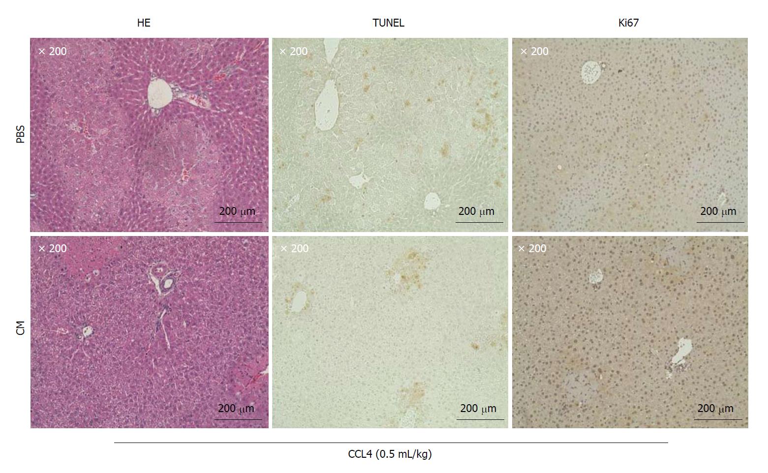
Figure 3 Culture supernatant concentrate significantly improved the symptoms of acute liver failure caused by the administration of CCL4.
Micrographic images of Hematoxylin and Eosin (HE) staining (left panels), TUNEL assay (middle panels) and tissue immunostaining of Ki67 (right panels) of liver specimens. Microscopic images of liver specimens 20 h after the administration of PBS (upper panels) and CM (lower panels) via the mouse tail vein. Fragmented DNA generated in the process of apoptosis can be detected by the TUNEL (TdT-mediated UTP nick end labeling) method. Ki67 protein present in the nucleus of cells in G1, S, G2 and M cycles (cell growth phase) was detected using immunostaining to identify cells in the growth phase in liver tissue. It was also used to count the number of positively stained cells in images of TUNNEL-stained sections (× 200). The numbers of positively stained cells in the PBS and CM groups were 14.00 ± 4.54 and 8.25 ± 5.57, respectively (n = 4; P = 0.19). The numbers of cells with positively stained nuclei on images of Ki67-stained sections (× 200) were also counted. The numbers of cells with positively stained nuclei in the PBS and CM groups were 9.25 ± 7.61 and 116.25 ± 3.06, respectively (n = 4; P < 0.01).
- Citation: Nahar S, Nakashima Y, Miyagi-Shiohira C, Kinjo T, Toyoda Z, Kobayashi N, Saitoh I, Watanabe M, Noguchi H, Fujita J. Cytokines in adipose-derived mesenchymal stem cells promote the healing of liver disease. World J Stem Cells 2018; 10(11): 146-159
- URL: https://www.wjgnet.com/1948-0210/full/v10/i11/146.htm
- DOI: https://dx.doi.org/10.4252/wjsc.v10.i11.146


