Copyright
©The Author(s) 2018.
World J Stem Cells. Nov 26, 2018; 10(11): 146-159
Published online Nov 26, 2018. doi: 10.4252/wjsc.v10.i11.146
Published online Nov 26, 2018. doi: 10.4252/wjsc.v10.i11.146
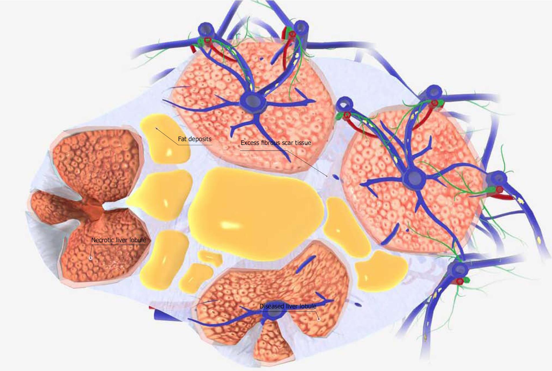
Figure 2 Schematic diagram of a liver cirrhosis tissue model.
The functional portion of the liver tissue is organized into hexagonal columns called liver lobules. Each liver lobule contains hundreds of individual liver cells (hepatocytes). In healthy liver tissue, adjacent lobules are separated by thin fibrous septa. However, liver cirrhosis involves thickening of the fibrous septa that separate lobules, and the deposition of fat. As a result, the blood flow in the lobules is disturbed and hepatocyte necrosis occurs. Also, the fibrous septa that separate the lobules transform the lobules and produce pseudolobules. Although it has a wide variety of causes, liver cirrhosis is most commonly caused by chronic alcohol abuse, chronic hepatitis, and nonalcoholic fatty liver disease. Images were obtained from BIODIGITAL HUMAN 3.0 (https://human.biodigital.com/index.html) (BioDigital, Broadway, NY, United States).
- Citation: Nahar S, Nakashima Y, Miyagi-Shiohira C, Kinjo T, Toyoda Z, Kobayashi N, Saitoh I, Watanabe M, Noguchi H, Fujita J. Cytokines in adipose-derived mesenchymal stem cells promote the healing of liver disease. World J Stem Cells 2018; 10(11): 146-159
- URL: https://www.wjgnet.com/1948-0210/full/v10/i11/146.htm
- DOI: https://dx.doi.org/10.4252/wjsc.v10.i11.146


