Copyright
©The Author(s) 2021.
World J Stem Cells. Aug 26, 2021; 13(8): 1084-1093
Published online Aug 26, 2021. doi: 10.4252/wjsc.v13.i8.1084
Published online Aug 26, 2021. doi: 10.4252/wjsc.v13.i8.1084
Figure 1 Availability of adipose-derived stem cells in patients after radiation exposure.
A: The number of stem cells harvested and growth rates did not appear to be affected by radiation; B: Decreased expression of endothelial markers of CD31, von Willebrand factor, and endothelial nitric oxide synthase between the adipose-derived stem cells from radiated (orange) and non-radiated (blue) breast tissue specimens. vWF: von Willebrand factor; eNOS: Endothelial nitric oxide synthase; ASC: Adipose-derived stem cell.
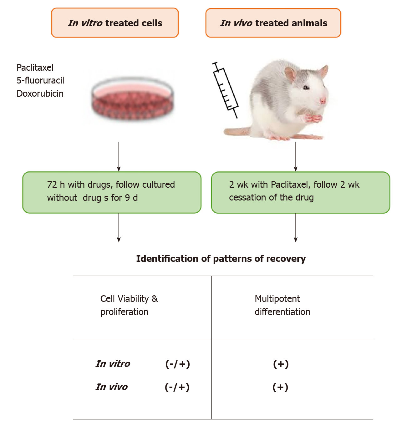
Figure 2 In vitro and in vivo evaluation of recovery potency of adipose-derived stem cells after chemotherapy exposure.
In both the in vitro and animal studies, after cessation of drugs, adipose-derived stem cells exhibited partial recovery (-/+) of cell growth and recovery (+) of multipotent differentiation capabilities.
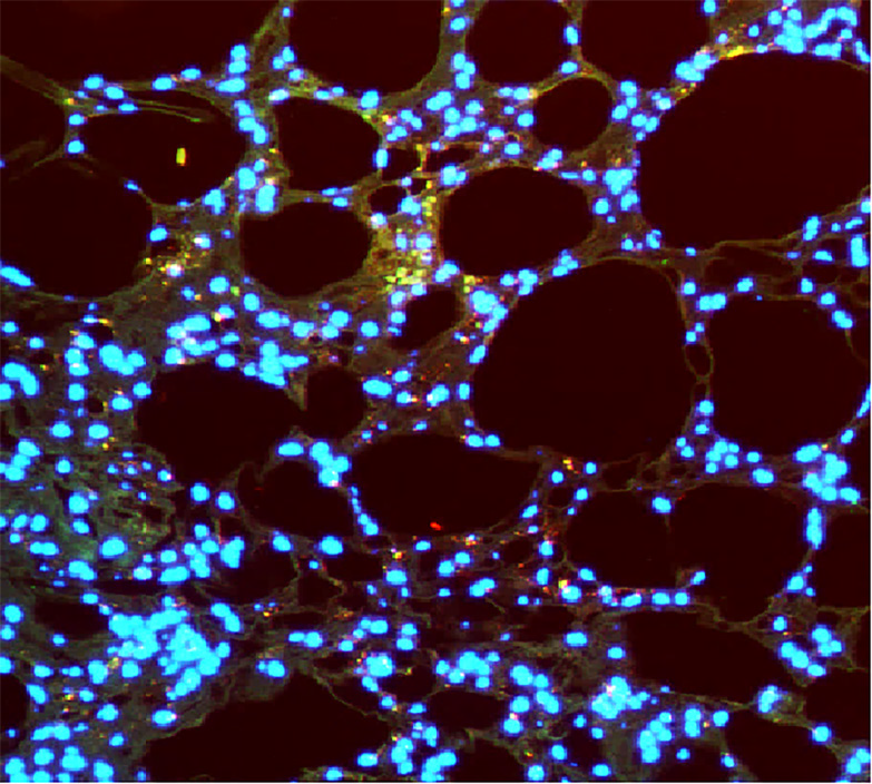
Figure 3 Adipose-derived stem cell-assisted transplanted fat lipoaspirate improved fat graft angiogenesis.
Immunofluorescence micrograph (200 ×; CD31 and human nuclear stain) of human adipose-derived stem cells (ASCs) with fat lipoaspirate injected into the rat for 8 wk. Fat lipoaspirate was mixed with human endothelial differentiated ASCs and then subcutaneously injected into the adult male Sprague-Dawley rat’s dorsum. Immunofluorescence staining analysis of the transplants was performed with an anti-human nuclear antibody (red) to detect if the human ASCs proliferated in transplanted tissues. The CD31 staining (green) was used to detect capillary endothelial cells. The merging of the red fluorescence of anti-human nuclear with the green fluorescence of CD31 revealed 3 yellow endothelial cells, indicating that the delivery of human ASCs promoted neovascularization.
- Citation: Platoff R, Villalobos MA, Hagaman AR, Liu Y, Matthews M, DiSanto ME, Carpenter JP, Zhang P. Effects of radiation and chemotherapy on adipose stem cells: Implications for use in fat grafting in cancer patients. World J Stem Cells 2021; 13(8): 1084-1093
- URL: https://www.wjgnet.com/1948-0210/full/v13/i8/1084.htm
- DOI: https://dx.doi.org/10.4252/wjsc.v13.i8.1084


