
















©The Author(s) 2003.
World J Gastroenterol. Dec 15, 2003; 9(12): 2751-2758
Published online Dec 15, 2003. doi: 10.3748/wjg.v9.i12.2751
Published online Dec 15, 2003. doi: 10.3748/wjg.v9.i12.2751
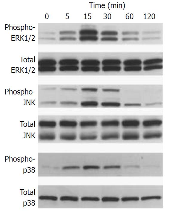
Figure 6 IL-1β activated MAP kinases.
SIPS cells were treated with IL-1β (at 2 ng/ml) for the indicated time. Total cell ly-sates (approximately 100 μg) were prepared, and the levels of activated, phosphorylated MAP kinases were determined by Western blotting. The levels of total MAP kinases were also determined.
- Citation: Masamune A, Satoh M, Kikuta K, Suzuki N, Shimosegawa T. Establishment and characterization of a rat pancreatic stellate cell line by spontaneous immortalization. World J Gastroenterol 2003; 9(12): 2751-2758
- URL: https://www.wjgnet.com/1007-9327/full/v9/i12/2751.htm
- DOI: https://dx.doi.org/10.3748/wjg.v9.i12.2751