
















©The Author(s) 2001.
World J Gastroenterol. Feb 15, 2001; 7(1): 16-21
Published online Feb 15, 2001. doi: 10.3748/wjg.v7.i1.16
Published online Feb 15, 2001. doi: 10.3748/wjg.v7.i1.16
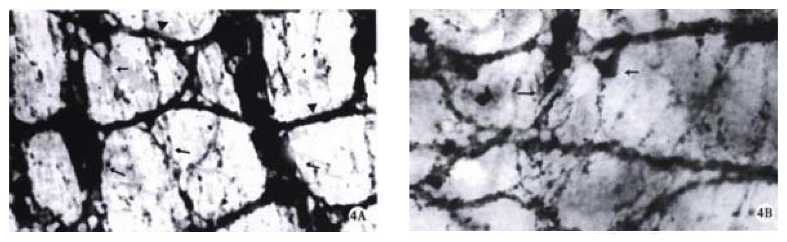
Figure 4 At ED 18, the myenteric plexuses of trisomy 16 mice and their normal littermates in whole mount preparation.
× 250 A. The developed myenteric plexus with primary (long arrow), secondary (arrow) and tertiary (arrowhead) strands were displayed in the colon of normal littermates. The primary strands were composed of broad fiber bundles and numerous ganglia with longitudinally arranged meshes. B. The myenteric plexus of trisomy 16 mice was poor with less ganglia containing some neurons (arrow) at different staining.
- Citation: Li JC, Mi KH, Zhou JL, Busch L, Kuhnel W. The development of colon innervation in trisomy 16 mice and Hirschsprungs disease. World J Gastroenterol 2001; 7(1): 16-21
- URL: https://www.wjgnet.com/1007-9327/full/v7/i1/16.htm
- DOI: https://dx.doi.org/10.3748/wjg.v7.i1.16