
















©The Author(s) 1998.
World J Gastroenterol. Aug 15, 1998; 4(4): 343-347
Published online Aug 15, 1998. doi: 10.3748/wjg.v4.i4.343
Published online Aug 15, 1998. doi: 10.3748/wjg.v4.i4.343
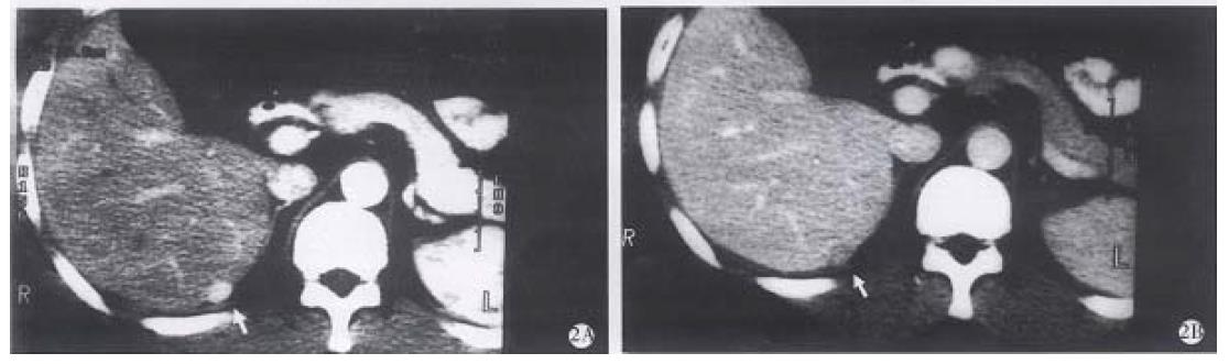
Figure 2 SHCC confirmed by surgery.
A.Arterial phase CT scan demonstrates the lesion enhanced homogenously and became hyperdense (arrow). B. Portal venous phase scan demonstrated the lesion was hypodense homogenously (arrow).
- Citation: Yan FH, Zeng MS, Zhou KR. Role and pitfalls of hepatic helical multi-phase CT scanning in differential diagnosis of small hemangioma and small hepatocellular carcinoma. World J Gastroenterol 1998; 4(4): 343-347
- URL: https://www.wjgnet.com/1007-9327/full/v4/i4/343.htm
- DOI: https://dx.doi.org/10.3748/wjg.v4.i4.343