
















Copyright: ©Author(s) 2026.
World J Gastroenterol. Jan 21, 2027; 33(3): 125206
Published online Jan 21, 2027. doi: 10.3748/wjg.125206
Published online Jan 21, 2027. doi: 10.3748/wjg.125206
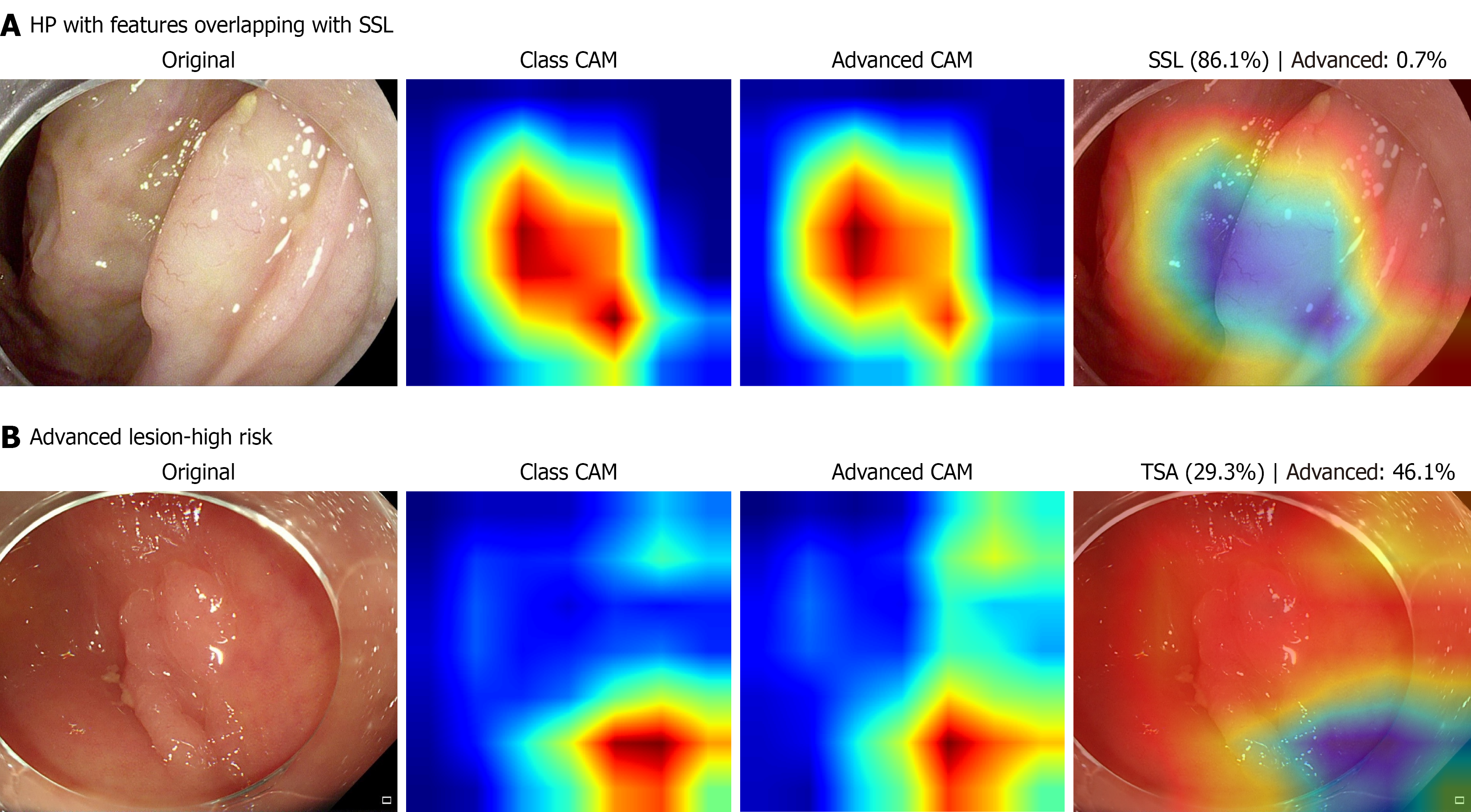
Figure 5 Gradient-weighted class activation mapping visualization of representative model predictions.
A: A hyperplastic polyp misclassified as a sessile serrated lesion, showing relatively increased gradient-weighted class activation mapping (Grad-CAM) activation over localized surface irregularities; B: A lesion with high-grade dysplasia correctly classified as high risk, showing focal Grad-CAM activation over the lesion region. For each case, the original white-light image, class activation map, high-grade dysplasia-risk activation map, and overlay image are shown from left to right. Warmer colors indicate areas with higher Grad-CAM activation. These visualizations are intended to provide qualitative insight into model behavior and should not be interpreted as definitive evidence of pathological relevance. HP: Hyperplastic polyp; SSL: Sessile serrated lesion; CAM: Class activation mapping; TSA: Traditional serrated adenoma.
- Citation: Zhang Y, Yan S, Li YJ, Li QX, Wei LJ. Deep learning for predicting high-grade dysplasia in colorectal polyps using white-light endoscopy. World J Gastroenterol 2027; 33(3): 125206
- URL: https://www.wjgnet.com/1007-9327/full/v33/i3/125206.htm
- DOI: https://dx.doi.org/10.3748/wjg.125206