
















Copyright: ©Author(s) 2026.
World J Gastroenterol. Jan 21, 2027; 33(3): 124453
Published online Jan 21, 2027. doi: 10.3748/wjg.124453
Published online Jan 21, 2027. doi: 10.3748/wjg.124453
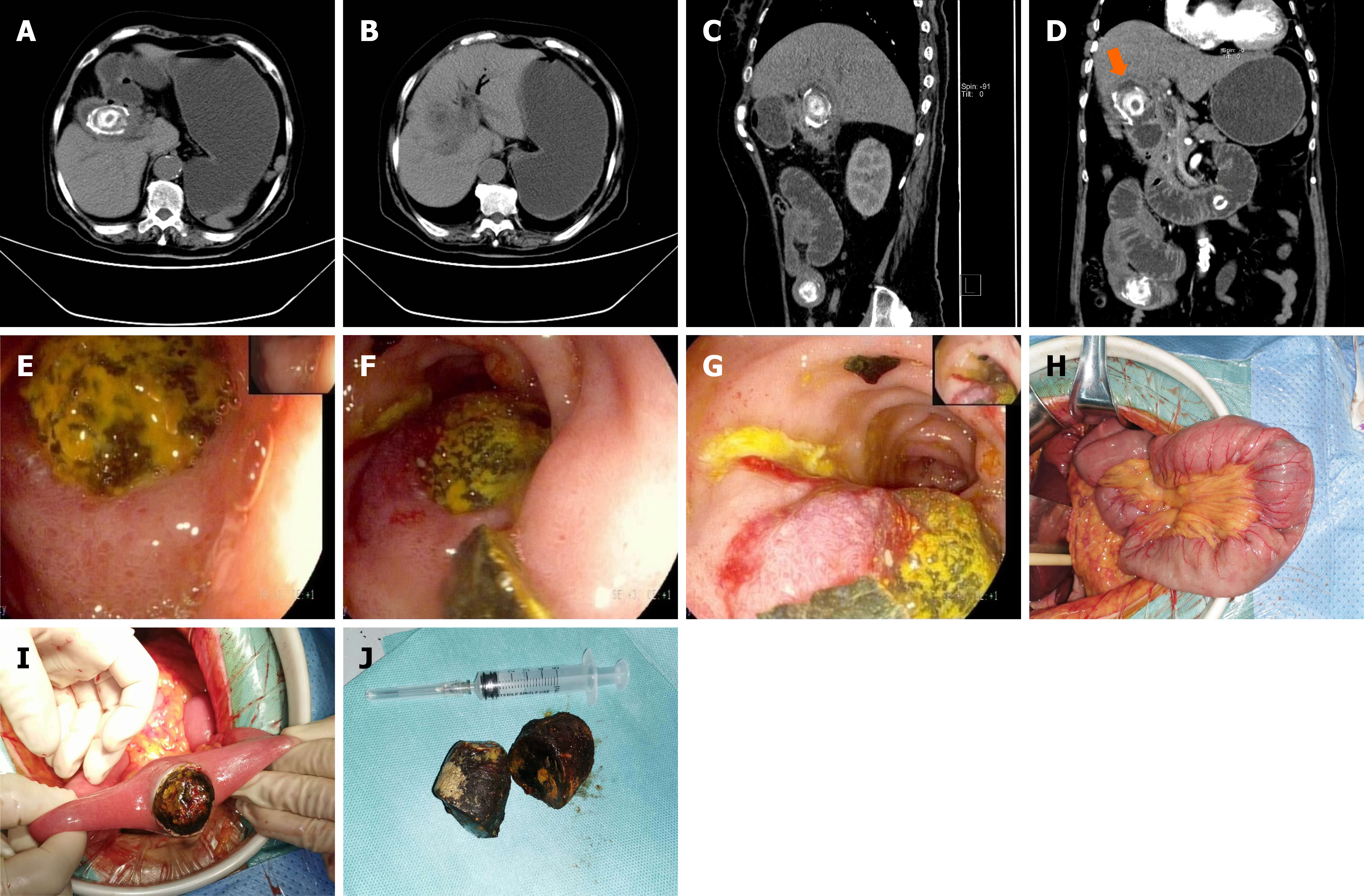
Figure 1 Imaging, endoscopic, intraoperative, and gross findings in a 72-year-old woman with Bouveret syndrome.
A: Axial abdominal computed tomography (CT) image demonstrating marked wall thickening in the duodenal papillary region and an annular, high-density intraluminal lesion corresponding to the impacted gallstone; B: Axial CT image demonstrating pneumobilia and dilated intrahepatic bile ducts; C: Sagittal CT image revealing two separate high-density intraluminal lesions, one in the duodenal bulb and the other in the small bowel, accompanied by proximal small-bowel dilatation; D: Coronal CT image demonstrating the impacted duodenal gallstone (orange arrow) and a second ectopic stone in the dilated jejunum; E: Enteroscopic image revealing a solitary spherical gallstone (diameter: About 5 cm) firmly lodged at the junction between the duodenal bulb and descending duodenum; F: Enteroscopic image demonstrating the rigidity of the gallstone, which could not be displaced through manipulation with biopsy forceps; G: Enteroscopic image showing the gallstone lodged at the junction between the duodenal bulb and descending duodenum, with surrounding mucosal hyperemia and edematous protrusion; H: Intraoperative photograph demonstrating dilatation of the proximal small bowel and the impacted bowel segment; I: Intraoperative photograph depicting removal of the second mobile gallstone from the jejunum, about 130 cm distal to the ligament of Treitz; J: Gross specimen image displaying the two extracted gallstones (each about 4 × 5 cm in size), both with faceted and laminated surfaces. A 5 mL syringe is shown for scale.
- Citation: Liu MX, Liu H. Bouveret syndrome with a synchronous jejunal gallstone in an elderly patient with high cardiac risk: A case report. World J Gastroenterol 2027; 33(3): 124453
- URL: https://www.wjgnet.com/1007-9327/full/v33/i3/124453.htm
- DOI: https://dx.doi.org/10.3748/wjg.124453