
















Copyright: ©Author(s) 2026.
World J Gastroenterol. Jan 21, 2027; 33(3): 124434
Published online Jan 21, 2027. doi: 10.3748/wjg.124434
Published online Jan 21, 2027. doi: 10.3748/wjg.124434
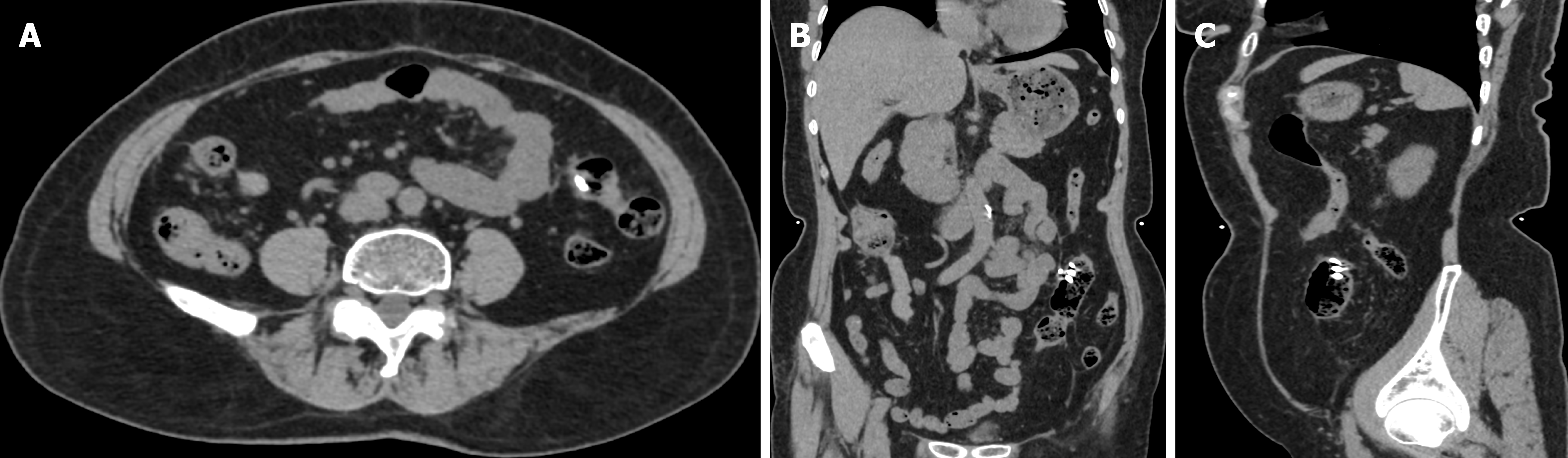
Figure 3 Abdominal computed tomography on postoperative day 35.
A-C: Axial, coronal, and sagittal views demonstrate several metallic clips at the repair site in the upper sigmoid colon, with mild localized wall thickening and intact serosa but no significant luminal narrowing; the pericolonic fat appears clear. Compared with the computed tomography obtained on postoperative day 1, fewer metallic clips are present, wall thickening and pericolonic fat stranding have markedly improved, the lumen remains patent, and the previously noted free air has completely resolved.
- Citation: Zeng X, Gong FR, Li Q, Shi YB, Lin F. Endoscopic closure of a giant iatrogenic sigmoid perforation using through-the-scope clips and a nylon loop: A case report. World J Gastroenterol 2027; 33(3): 124434
- URL: https://www.wjgnet.com/1007-9327/full/v33/i3/124434.htm
- DOI: https://dx.doi.org/10.3748/wjg.124434