
















Copyright: ©Author(s) 2026.
World J Gastroenterol. Jan 21, 2027; 33(3): 123466
Published online Jan 21, 2027. doi: 10.3748/wjg.123466
Published online Jan 21, 2027. doi: 10.3748/wjg.123466
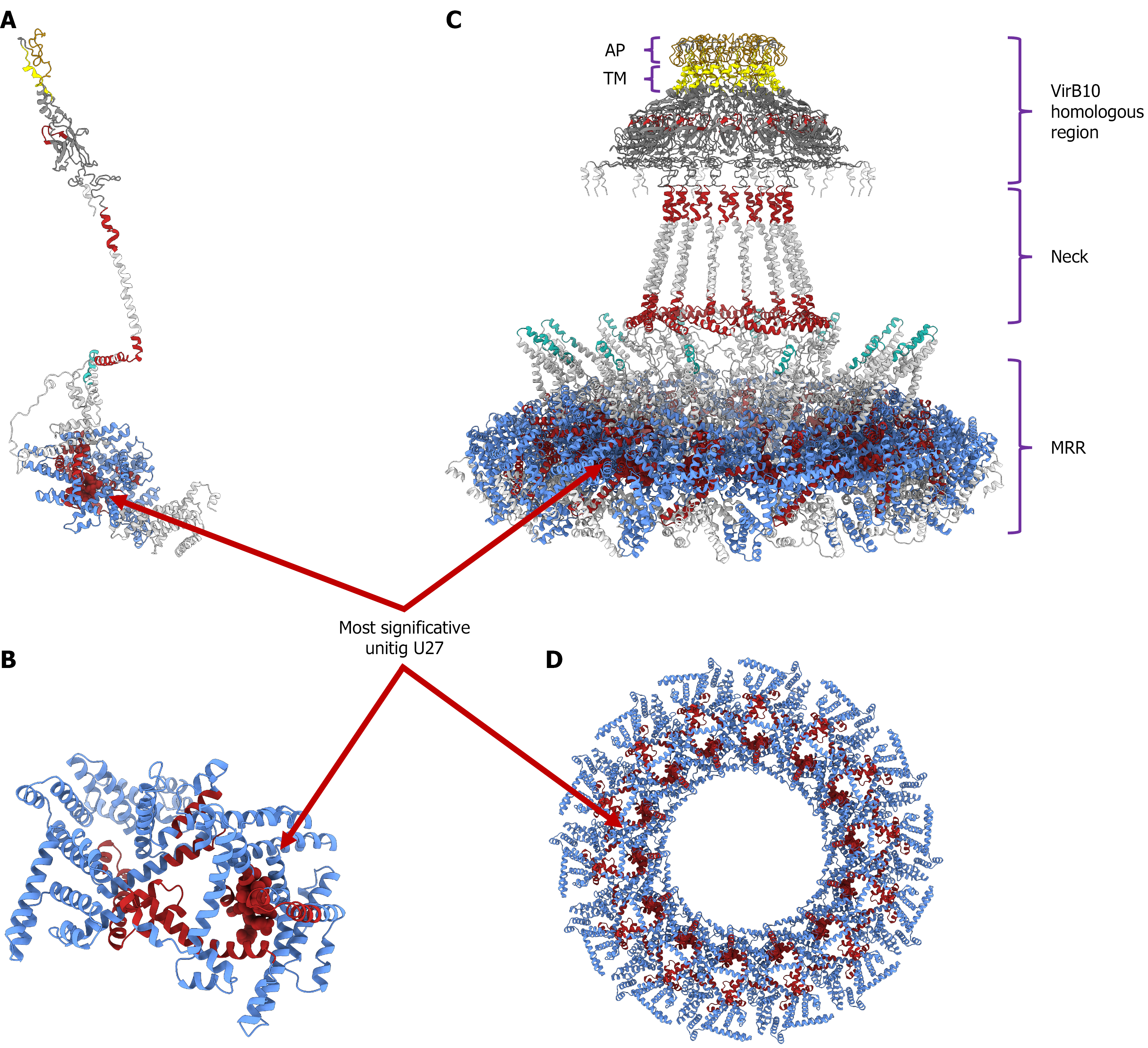
Figure 6 Three-dimensional structural models of the cagY protein (strain 26695) and disease-associated regions.
Structural domains and clinical associations are color-coded as follows: Middle repeat region (MRR) (blue), cagY backbone (white), transmembrane region (TM) (yellow), non-atrophic gastritis-associated regions (turquoise), and gastric cancer-associated regions (red). A: Structural model of the cagY monomer, showing the spatial distribution of significant phenotype-associated regions (P < 0.01); B: Close-up view of the monomeric MRR, highlighting the specific locus of the most significant unitig (U27); C: Lateral view of the multimeric cagY complex assembly, detailing the vertical organization of the VirB10 homologous region comprising the antenna projection and TM domains, the neck, and the base MRR; D: Axial (top-down) view of the multimeric MRR tunnel, showing the ring-like distribution and exposure of the most significant unitig (U27). AP: Antenna projection; TM: Transmembrane region; MRR: Middle repeat region.
- Citation: López-Luis MA, Torres RC, Solnick JV, Camargo MC, Méndez-Tenorio A, Torres J. In-depth analysis of cagY in worldwide Helicobacter pylori strains unmasks a strong correlation between sequence diversity and gastric cancer. World J Gastroenterol 2027; 33(3): 123466
- URL: https://www.wjgnet.com/1007-9327/full/v33/i3/123466.htm
- DOI: https://dx.doi.org/10.3748/wjg.123466