
















Copyright: ©Author(s) 2026.
World J Gastroenterol. Jan 14, 2027; 33(2): 125781
Published online Jan 14, 2027. doi: 10.3748/wjg.125781
Published online Jan 14, 2027. doi: 10.3748/wjg.125781
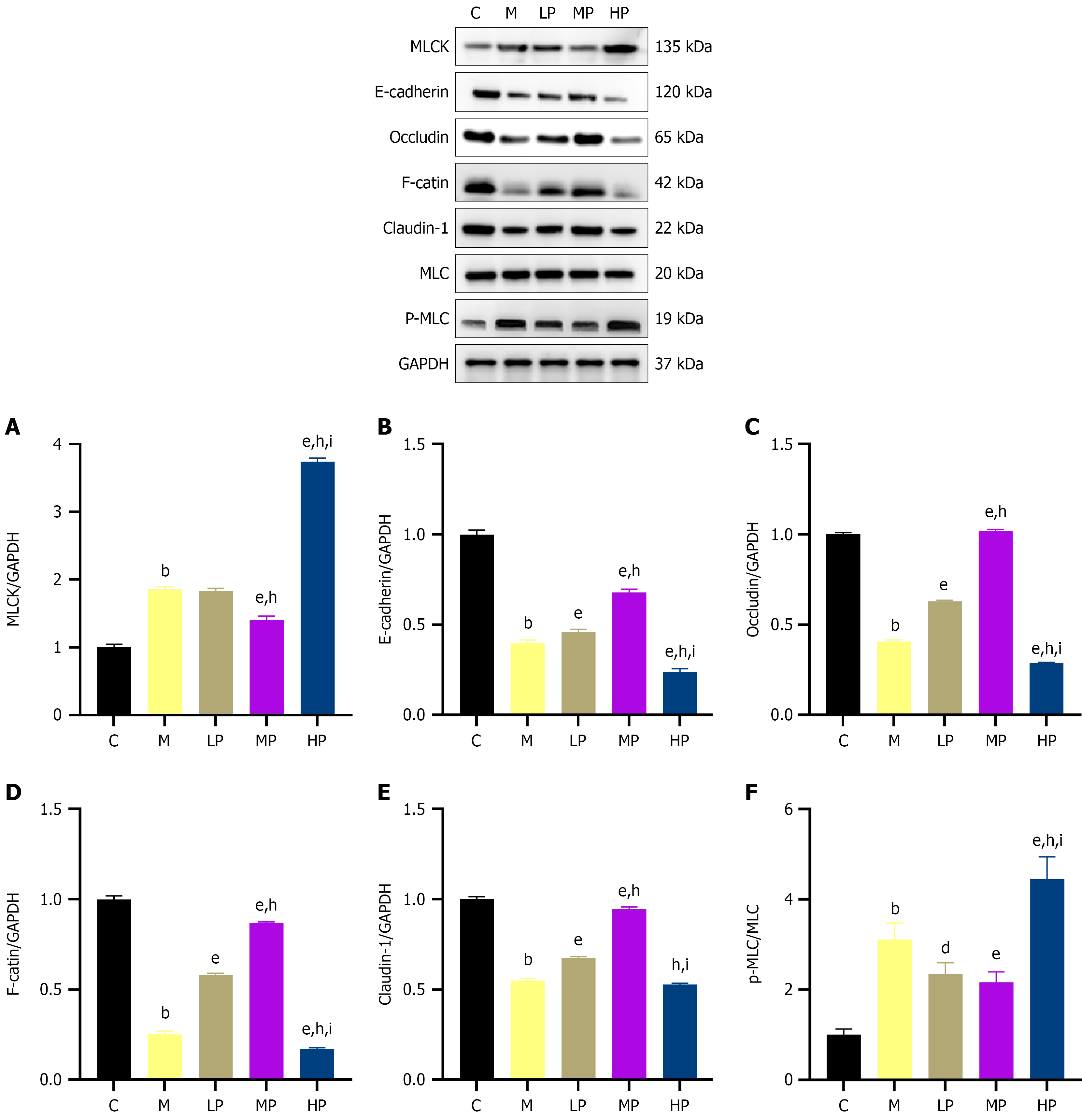
Figure 12 Effect of massage on the myosin light chain kinase/phosphorylated myosin light chain/E-cadherin signaling axis and epithelial barrier-related protein expression in duodenal tissue of rabbits with functional dyspepsia (n = 6).
bP < 0.001 vs the control group; dP < 0.01 vs the model group; eP < 0.001 vs the model group; hP < 0.001 vs the light-pressure group; iP < 0.001 vs the medium-pressure group. A-F: Representative western blot bands show protein expression of myosin light chain kinase, E-cadherin, Occludin, F-actin, Claudin-1, myosin light chain, phosphorylated myosin light chain, and glyceraldehyde-3-phosphate dehydrogenase in duodenal tissue. MLC: Myosin light chain; p-MLC: Phosphorylated myosin light chain; MLCK: Myosin light chain kinase; GAPDH: Glyceraldehyde-3-phosphate dehydrogenase; C: The control group; M: The model group; LP: The light-pressure group; MP: The medium-pressure group; HP: The heavy-pressure group.
- Citation: Zhang YC, Yang WX, Hou JL, Wang ZX, Huang ZK, Li HL, Kek MZ, Lin DR, Wu RH, Wang JH. Robot-assisted massage is associated with improved duodenal barrier-related outcomes in a rabbit model of functional dyspepsia. World J Gastroenterol 2027; 33(2): 125781
- URL: https://www.wjgnet.com/1007-9327/full/v33/i2/125781.htm
- DOI: https://dx.doi.org/10.3748/wjg.125781