
















Copyright: ©Author(s) 2026.
World J Gastroenterol. Jan 14, 2027; 33(2): 124537
Published online Jan 14, 2027. doi: 10.3748/wjg.124537
Published online Jan 14, 2027. doi: 10.3748/wjg.124537
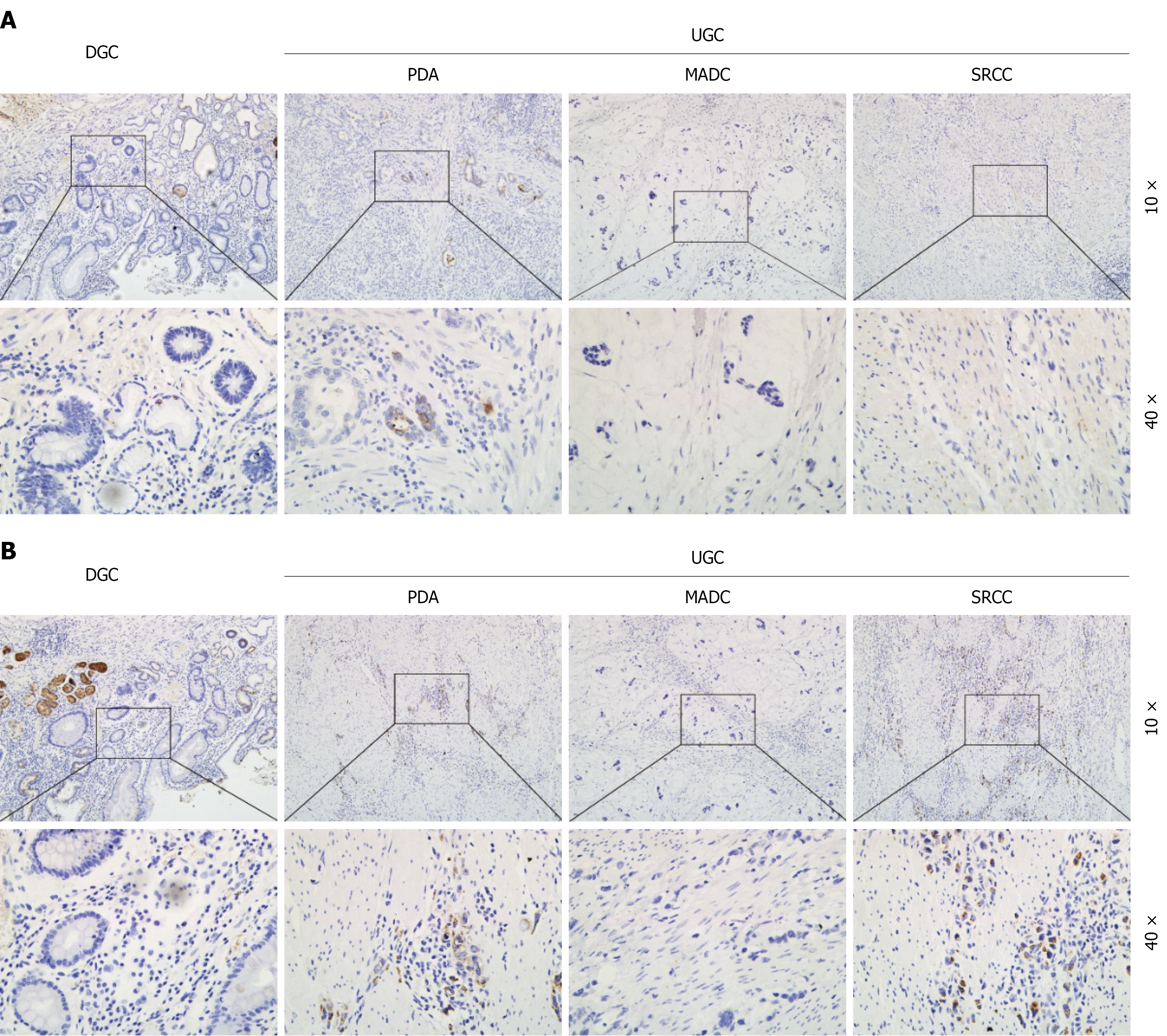
Figure 3 Representative immunohistochemical staining for pepsinogen in different histological types of gastric cancer.
A: Pepsinogen (PG) I staining; B: PG II staining. Positive immunoreactivity appeared as brown cytoplasmic staining. Original magnification: 10 × and 40 × (objective). DGC: Differentiated gastric cancer; UGC: Undifferentiated gastric cancer; PDA: Poorly differentiated adenocarcinoma; MADC: Mucinous adenocarcinoma; SRCC: Signet ring cell carcinoma.
- Citation: Zhang JR, Xu XW, Liu JM, Zhu ZH, Mao CM. Serum pepsinogen combined with computed tomography imaging for diagnosing recurrence of gastric cancer after total gastrectomy. World J Gastroenterol 2027; 33(2): 124537
- URL: https://www.wjgnet.com/1007-9327/full/v33/i2/124537.htm
- DOI: https://dx.doi.org/10.3748/wjg.124537