










Copyright: ©Author(s) 2026.
World J Gastroenterol. Jan 14, 2027; 33(2): 124537
Published online Jan 14, 2027. doi: 10.3748/wjg.124537
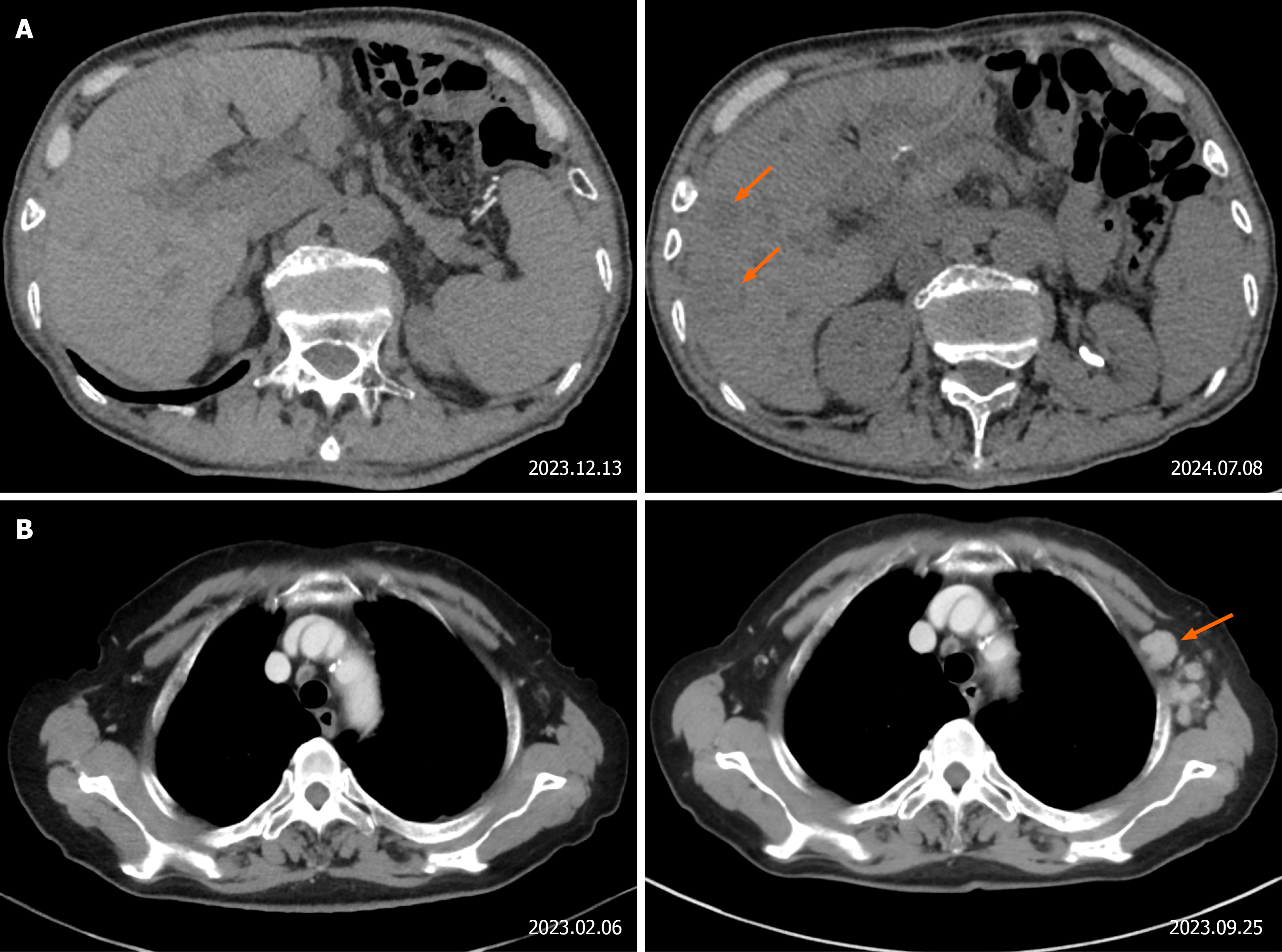
Figure 1 Representative cases with elevated serum pepsinogen levels in the early postoperative period and negative concurrent com puted tomography imaging, with recurrence subsequently confirmed on follow-up imaging.
A: A 73-year-old male with an early postoperative serum pepsinogen (PG) I concentration of 30.97 ng/mL and a PG II concentration of 2.25 ng/mL. Concurrent computed tomography (CT) imaging revealed no evidence of metastasis. At 6 months post-surgery, CT imaging showed multiple liver metastases (orange arrows); B: An 80-year-old female with an early postoperative serum PG I concentration of 27.10 ng/mL and a PG II concentration of 1.83 ng/mL. Concurrent CT imaging was negative for metastasis. At 7 months post-surgery, CT imaging revealed widespread lymph node metastases, including left axillary lymph node metastasis (orange arrow).
- Citation: Zhang JR, Xu XW, Liu JM, Zhu ZH, Mao CM. Serum pepsinogen combined with computed tomography imaging for diagnosing recurrence of gastric cancer after total gastrectomy. World J Gastroenterol 2027; 33(2): 124537
- URL: https://www.wjgnet.com/1007-9327/full/v33/i2/124537.htm
- DOI: https://dx.doi.org/10.3748/wjg.124537