
















Copyright: ©Author(s) 2027.
World J Gastroenterol. Jan 14, 2027; 33(2): 123953
Published online Jan 14, 2027. doi: 10.3748/wjg.123953
Published online Jan 14, 2027. doi: 10.3748/wjg.123953
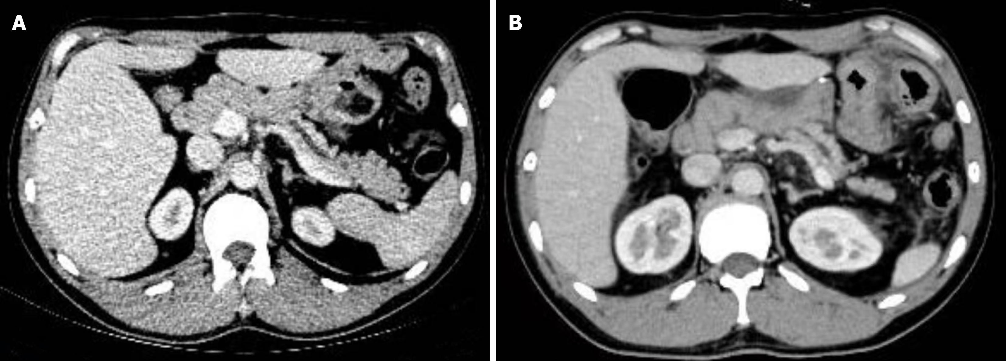
Figure 4 Case 2 follow-up contrast-enhanced computed tomography.
A: Axial image at 12 months; B: Axial image at 24 months. Both demonstrate expected postoperative changes and mild dilatation of the main pancreatic duct.
- Citation: Zhang YP, Lu Y, Wei HZ, Yang M, Liang B, He L, Zhao CB, Deng ZL, Wang JJ, Wang GQ, Wu HS, Yin T. Gastroduodenal arterioportal fistula: Two case reports and review of literature. World J Gastroenterol 2027; 33(2): 123953
- URL: https://www.wjgnet.com/1007-9327/full/v33/i2/123953.htm
- DOI: https://dx.doi.org/10.3748/wjg.123953