
















Copyright: ©Author(s) 2027.
World J Gastroenterol. Jan 14, 2027; 33(2): 123953
Published online Jan 14, 2027. doi: 10.3748/wjg.123953
Published online Jan 14, 2027. doi: 10.3748/wjg.123953
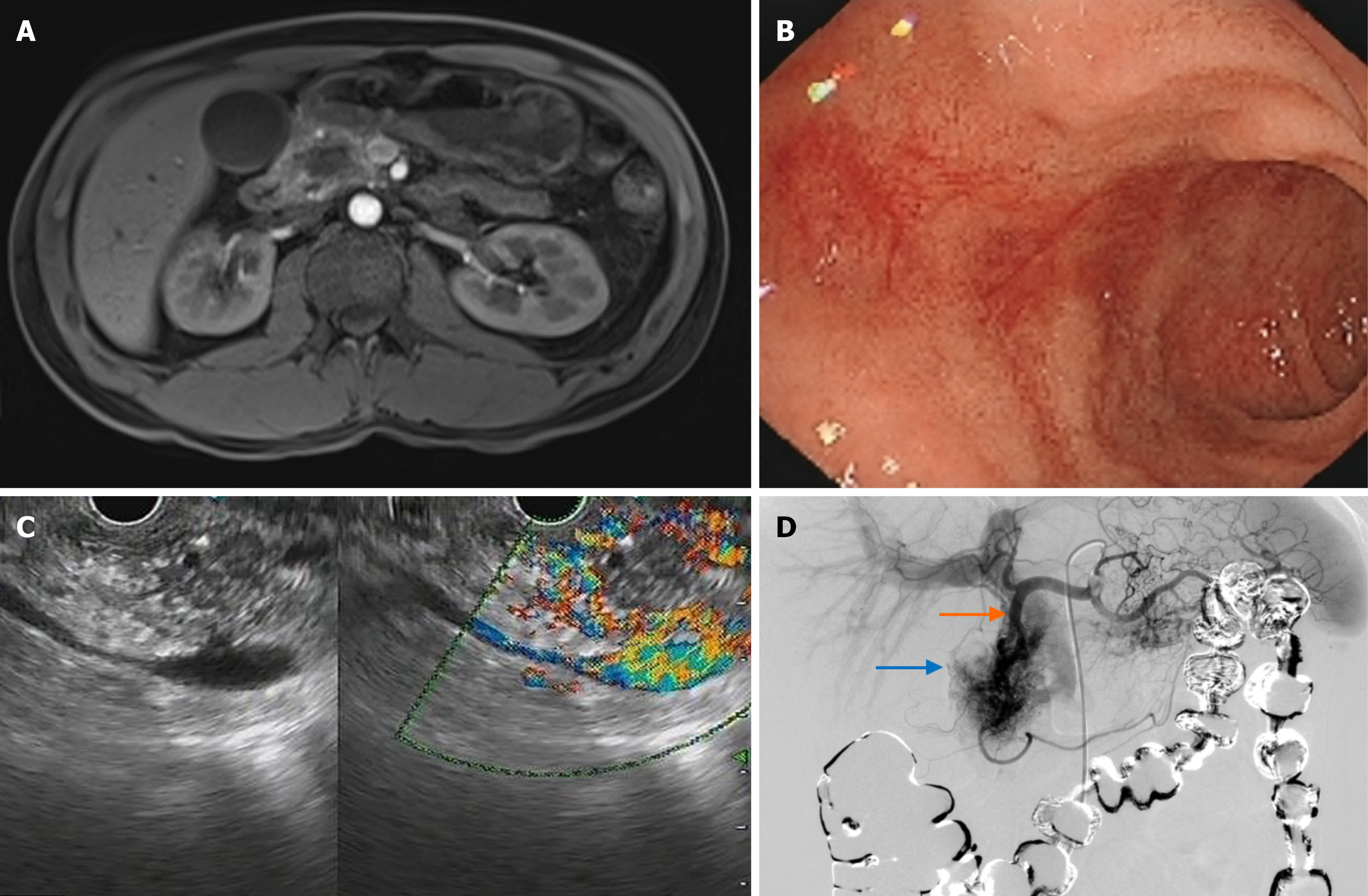
Figure 2 Case 2 preoperative imaging and angiography.
A: Magnetic resonance cholangiopancreatography showing a 21 mm × 14 mm T1-hypointense lesion in the pancreatic head; B: Esophagogastroduodenoscopy revealed fresh blood adherent to the mucosa at the junction of the duodenal bulb and descending duodenum, without an identifiable ulcer or neoplastic lesion; C: Endoscopic ultrasonography demonstrating a well-defined hypoechoic mass in the uncinate process with prominent peripheral blood flow on color Doppler; D: Arteriography demonstrating the abnormal vascular network in the descending gastroduodenal artery (blue arrow) and the gastroduodenal artery (orange arrow).
- Citation: Zhang YP, Lu Y, Wei HZ, Yang M, Liang B, He L, Zhao CB, Deng ZL, Wang JJ, Wang GQ, Wu HS, Yin T. Gastroduodenal arterioportal fistula: Two case reports and review of literature. World J Gastroenterol 2027; 33(2): 123953
- URL: https://www.wjgnet.com/1007-9327/full/v33/i2/123953.htm
- DOI: https://dx.doi.org/10.3748/wjg.123953