
















Copyright: ©Author(s) 2026.
World J Gastroenterol. Jan 14, 2027; 33(2): 123795
Published online Jan 14, 2027. doi: 10.3748/wjg.123795
Published online Jan 14, 2027. doi: 10.3748/wjg.123795
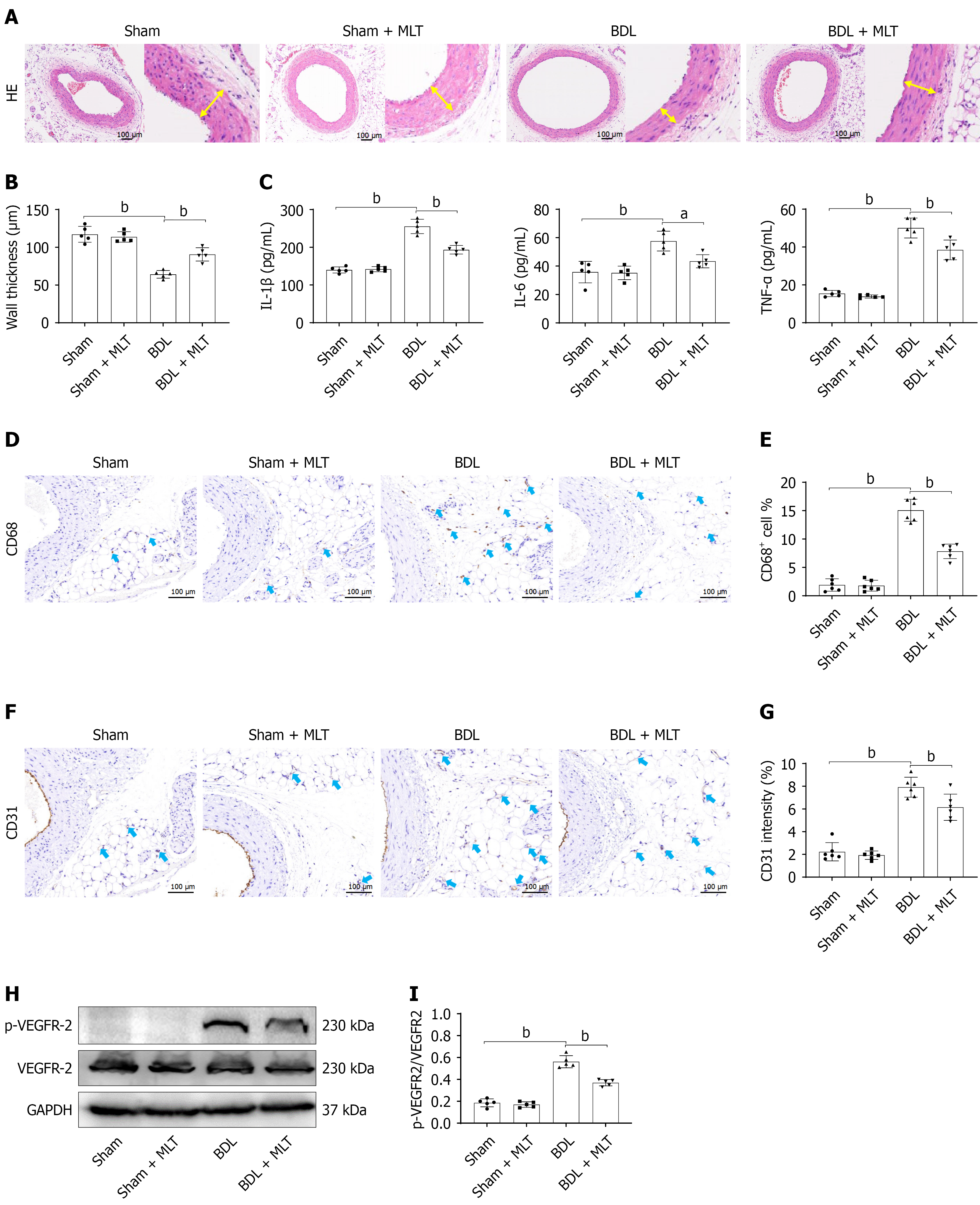
Figure 2 Melatonin alleviated bile duct ligation-induced mesenteric vascular remodeling and angiogenesis.
A: Representative hematoxylin and eosin staining images of mesenteric artery. The yellow arrows represent the thickness of vascular wall; B: Quantitative analysis of the wall thickness; C: Enzyme-linked immunosorbent assay of inflammatory cytokines in mesenteric artery tissues; D: Representative immunohistochemical images of CD68 in the mesentery; E: Quantitative analysis of CD68-positive cells; F: Representative immunohistochemical images of CD31 in the mesentery; G: Quantitative analysis of CD31 staining intensity; H: Western blotting of mesenteric VEGFR-2 and its phosphorylated form; I: Quantitative analysis of VEGFR-2 phosphorylation. aP < 0.05. bP < 0.01. BDL: Bile duct ligation; MLT: Melatonin; HE: Hematoxylin and eosin; IL: Interleukin; TNF-α: Tumor necrosis factor alpha.
- Citation: Wu GB, Luo GQ, Chen M, Cai KW, You XT, Han F, Zhang NZ, Luo M, Li Q, Huo HZ, Fan Q. Melatonin attenuates splanchnic hyperdynamic circulation in cirrhotic portal hypertension by disrupting POMK-VEGFR2 interaction. World J Gastroenterol 2027; 33(2): 123795
- URL: https://www.wjgnet.com/1007-9327/full/v33/i2/123795.htm
- DOI: https://dx.doi.org/10.3748/wjg.123795