
















Copyright: ©Author(s) 2027.
World J Gastroenterol. Jan 7, 2027; 33(1): 123843
Published online Jan 7, 2027. doi: 10.3748/wjg.123843
Published online Jan 7, 2027. doi: 10.3748/wjg.123843
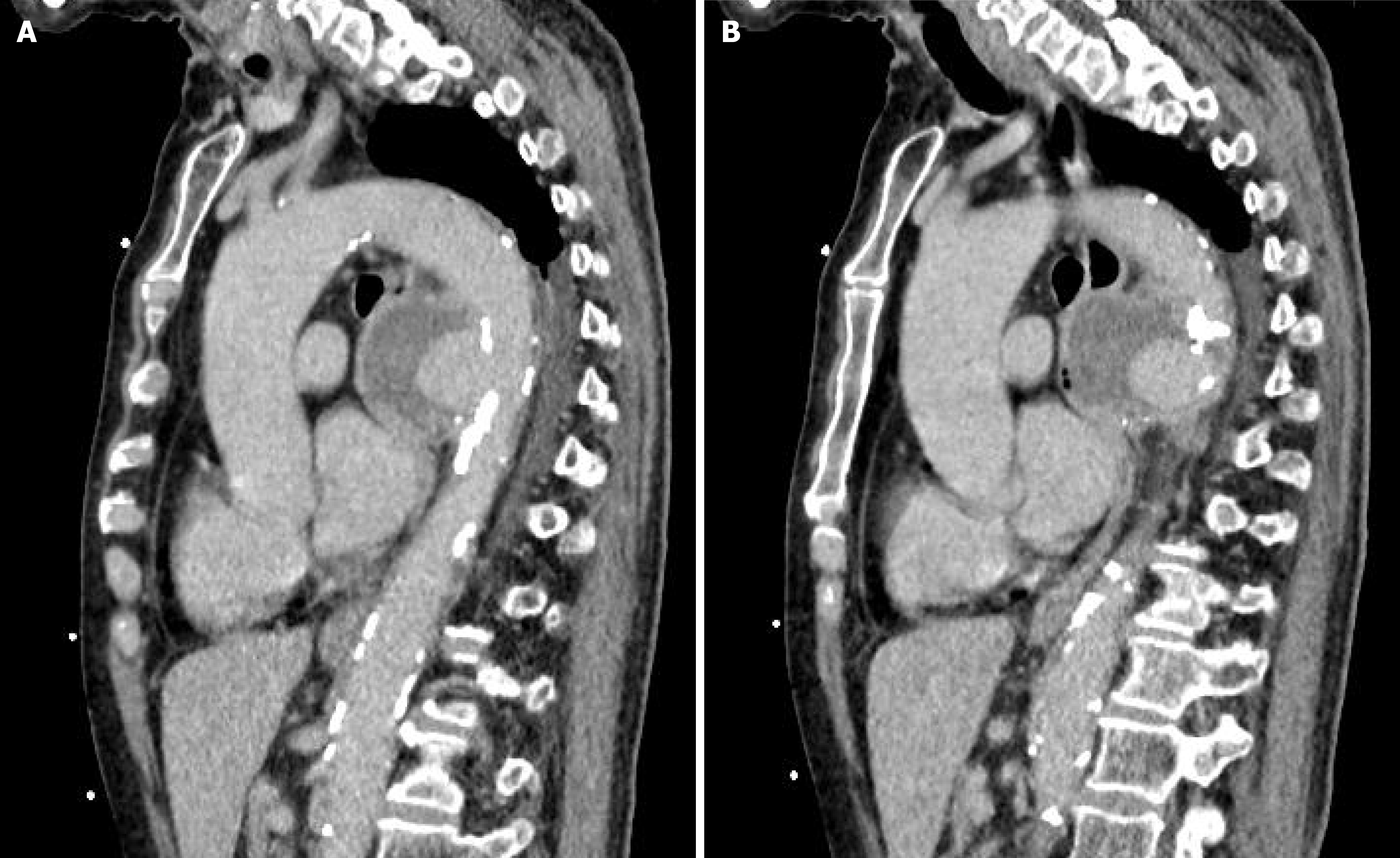
Figure 3 Sagittal computed tomography angiography of the descending thoracic aorta.
A: A sagittal image showing the saccular pseudoaneurysm with central contrast enhancement and peripheral low-attenuation components compatible with mural thrombus or perianeurysmal inflammatory tissue; B: An adjacent sagittal image showing compression and anterior displacement of the esophagus by the pseudoaneurysm.
- Citation: Xue Y, Yun XJ, Liu QY, Qi YT, Zhang LH, Jiang K, Li BD, Zhang JX. Suspected Brucella-associated descending thoracic aortic pseudoaneurysm presenting as upper gastrointestinal bleeding: A case report and review of literature. World J Gastroenterol 2027; 33(1): 123843
- URL: https://www.wjgnet.com/1007-9327/full/v33/i1/123843.htm
- DOI: https://dx.doi.org/10.3748/wjg.123843