
















Copyright: ©Author(s) 2027.
World J Gastroenterol. Jan 7, 2027; 33(1): 123681
Published online Jan 7, 2027. doi: 10.3748/wjg.123681
Published online Jan 7, 2027. doi: 10.3748/wjg.123681
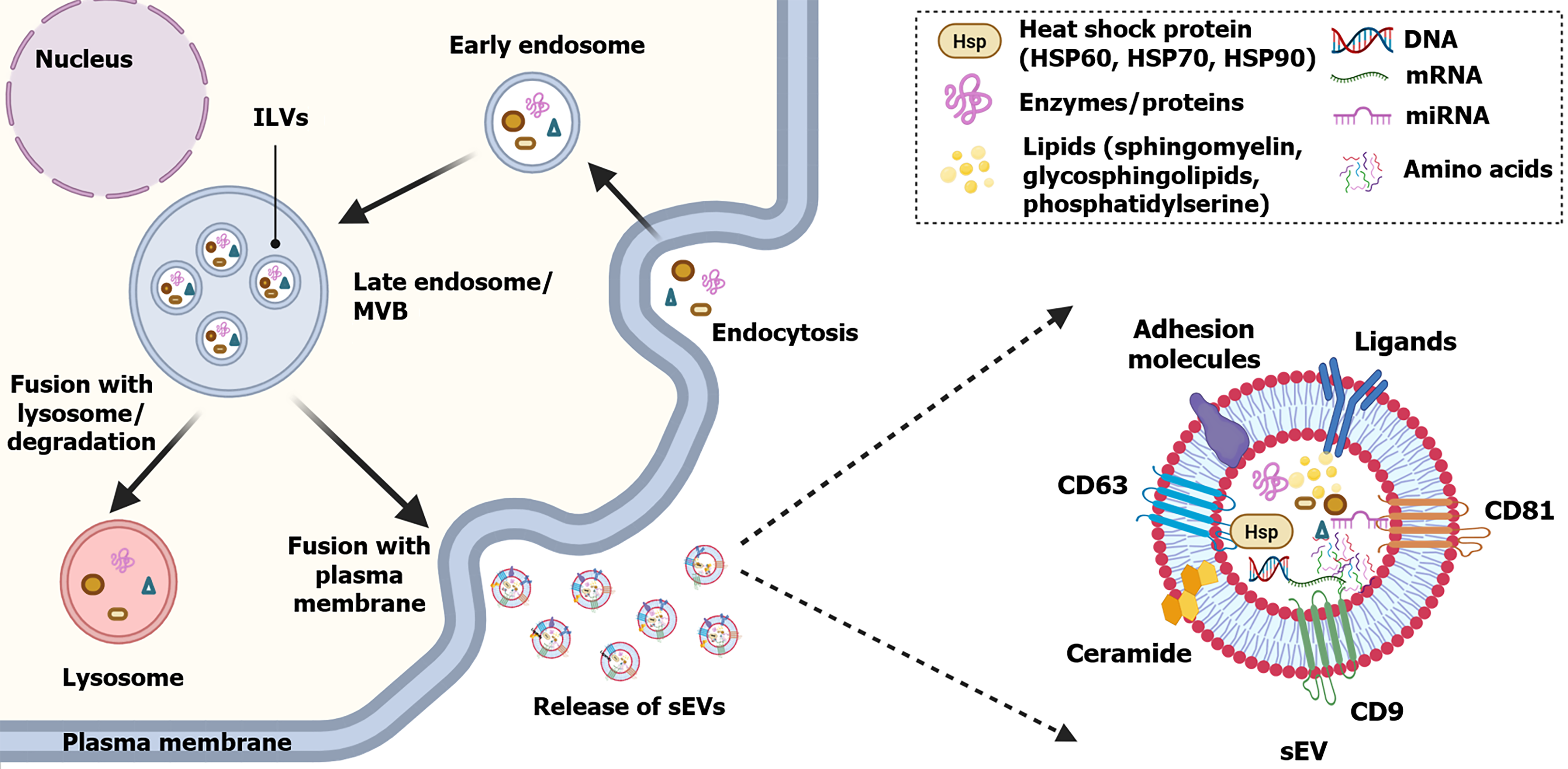
Figure 1 Conceptual schematic of small extracellular vesicles biogenesis and structure.
Endocytosis leads to the formation of early endosomes, followed by endosomal maturation and the generation of multivesicular bodies (MVBs). Intraluminal vesicles are formed by inward budding of the endosomal limiting membrane within MVBs. MVBs may either fuse with lysosomes for degradation or fuse with the plasma membrane, resulting in the extracellular release of intraluminal vesicles as small extracellular vesicles. Small extracellular vesicles contain diverse cargos, including nucleic acids, proteins, lipids, and enzymes, and may display extracellular vesicles (EV)-associated tetraspanins such as CD9, CD63, and CD81. These proteins are commonly used for EV characterization but are not universal or definitive markers of a specific EV subtype; multiparametric characterization is recommended. ILVs: Intraluminal vesicles; MVB: Multivesicular body; sEVs: Small extracellular vesicles; HSP: Heat shock protein.
- Citation: Ding LL, Liu H, Yu Y, Liu XC, Zhang SQ, Qian H, Hao DL. Small extracellular vesicles as liquid biopsy targets and precision therapeutics for metabolic dysfunction-associated steatotic liver disease. World J Gastroenterol 2027; 33(1): 123681
- URL: https://www.wjgnet.com/1007-9327/full/v33/i1/123681.htm
- DOI: https://dx.doi.org/10.3748/wjg.123681