
















©Author(s) (or their employer(s)) 2026.
World J Gastroenterol. Mar 7, 2026; 32(9): 114302
Published online Mar 7, 2026. doi: 10.3748/wjg.v32.i9.114302
Published online Mar 7, 2026. doi: 10.3748/wjg.v32.i9.114302
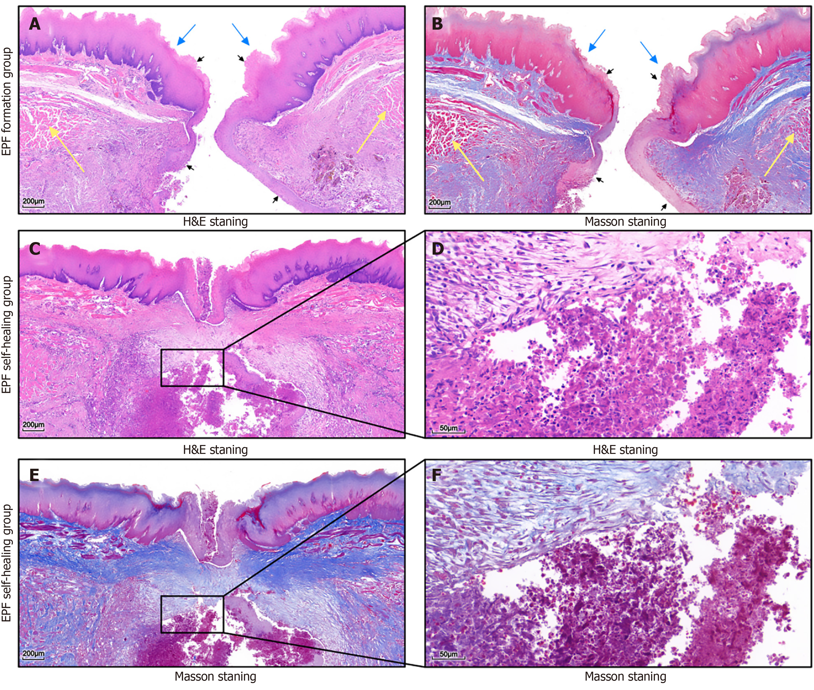
Figure 6 Histological observations.
A and B: Histological analysis shows disruption of the esophageal mucosa (blue arrow) and muscular layer (yellow arrow), with esophageal squamous epithelium extending towards the thoracic cavity (black arrow), representing the formation of esophageal pleural fistula (EPF); C-F: The EPF self-healing group shows that mucosal restitution is the primary reparative event; the abscess cavity contains abundant necrotic debris and infiltrating inflammatory cells, surrounded by newly synthesized fibrous tissue that initiates the encapsulation of the abscess. H&E: Hematoxylin & eosin; EPF: Esophageal pleural fistula.
- Citation: He Q, Chen XY, Xiang Q, Huang CH, Wu ZW. Establishing an esophago-pleural fistula disease model in rabbits with a magnetic compression technique. World J Gastroenterol 2026; 32(9): 114302
- URL: https://www.wjgnet.com/1007-9327/full/v32/i9/114302.htm
- DOI: https://dx.doi.org/10.3748/wjg.v32.i9.114302