
















©Author(s) (or their employer(s)) 2026.
World J Gastroenterol. Mar 7, 2026; 32(9): 114302
Published online Mar 7, 2026. doi: 10.3748/wjg.v32.i9.114302
Published online Mar 7, 2026. doi: 10.3748/wjg.v32.i9.114302
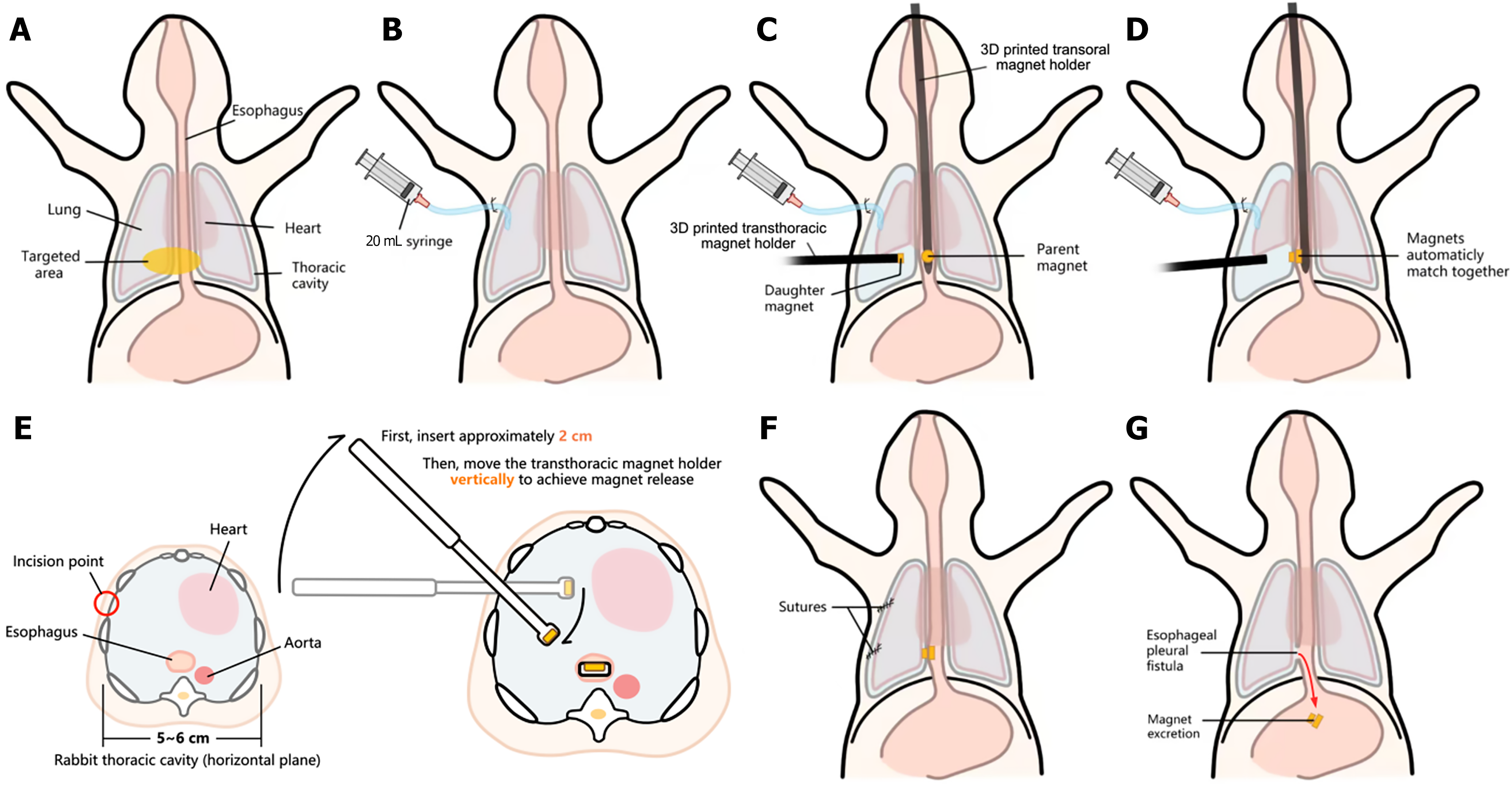
Figure 3 Schematic diagram of the esophageal pleural fistula modeling process.
A: The primary operative site (marked yellow); B: Insertion of a 20 mL syringe-connected catheter into the thoracic cavity; C: After injecting 20 mL of air to create space for operation, the transthoracic magnet holder and the transoral magnet holder were inserted; D: Magnetic force pulls the magnets together, compressing the tissue in between; E: A cross-sectional view at the xiphoid level of the thorax; F: Surgical closure after removal of both magnet holders; G: Magnets naturally fall off within 6-8 days postoperatively.
- Citation: He Q, Chen XY, Xiang Q, Huang CH, Wu ZW. Establishing an esophago-pleural fistula disease model in rabbits with a magnetic compression technique. World J Gastroenterol 2026; 32(9): 114302
- URL: https://www.wjgnet.com/1007-9327/full/v32/i9/114302.htm
- DOI: https://dx.doi.org/10.3748/wjg.v32.i9.114302