
















©Author(s) (or their employer(s)) 2026.
World J Gastroenterol. Mar 7, 2026; 32(9): 114207
Published online Mar 7, 2026. doi: 10.3748/wjg.v32.i9.114207
Published online Mar 7, 2026. doi: 10.3748/wjg.v32.i9.114207
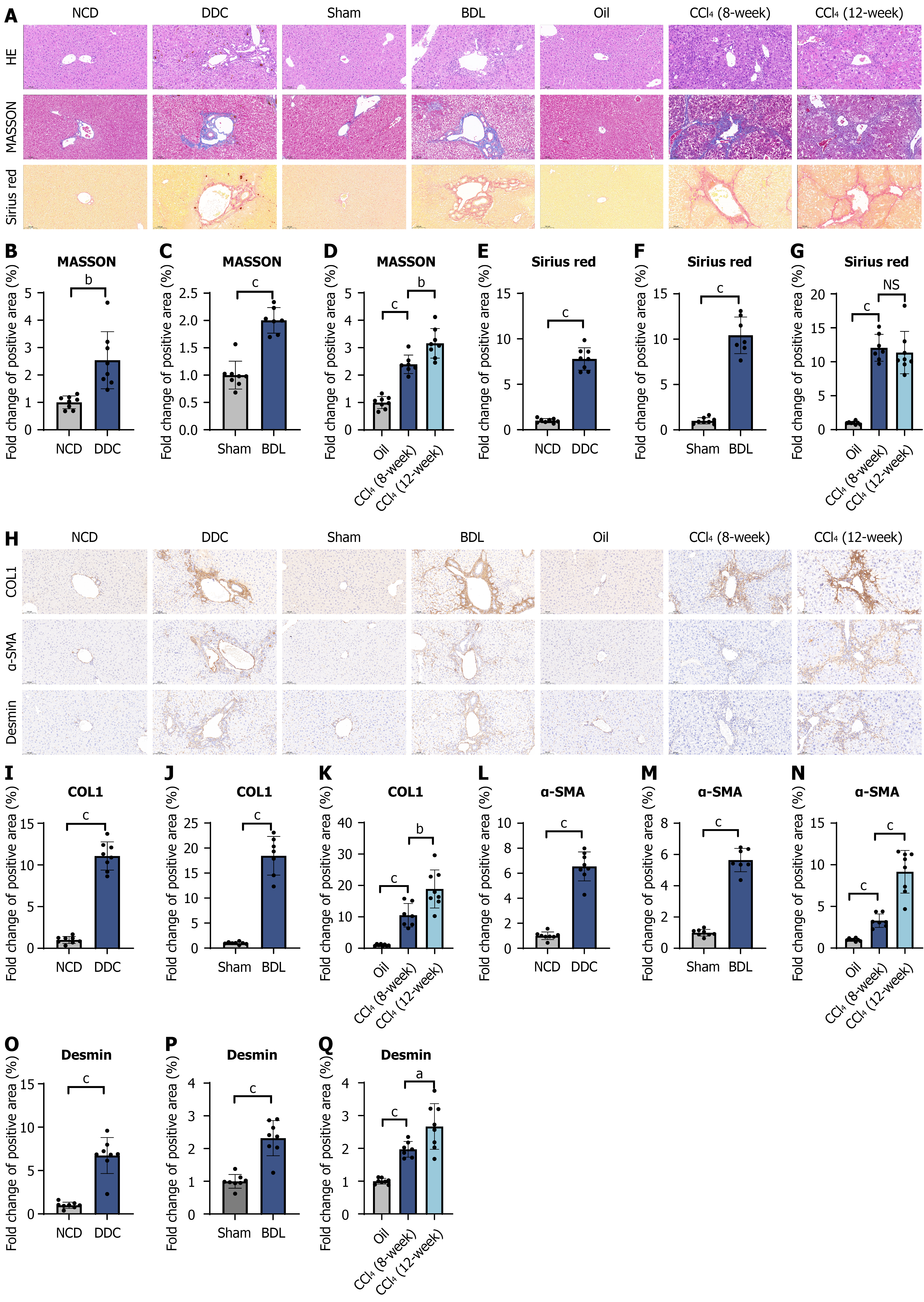
Figure 2 The 3,5-diethoxycarbonyl-1,4-dihydrocollidine diet mouse model (4 weeks) exhibited moderate biliary fibrosis in the portal vein region.
A: Hematoxylin-eosin, Masson, and Sirius red staining of mouse livers; B-G: Statistical analysis of Masson and Sirius red staining results; H: Immunohistochemistry staining for collagen-1, α-smooth muscle actin (SMA), and desmin in mouse livers; I-Q: Statistical analysis of collagen-1, α-SMA, and desmin staining results. Sample sizes (n): 8:8 (normal chow diet vs 3,5-diethoxycarbonyl-1,4-dihydrocollidine), 8:7 (sham vs bile duct ligation), and 8:7:8 (oil vs 8-week carbon tetrachloride vs 12-week carbon tetrachloride). aP < 0.05. bP < 0.01. cP < 0.001. NS: No significant; NCD: Normal chow diet; DDC: 3,5-diethoxycarbonyl-1,4-dihydrocollidine; BDL: Bile duct ligation; CCl4: Carbon tetrachloride; HE: Hematoxylin-eosin; COL1: Collagen-1; SMA: Smooth muscle actin.
- Citation: Zhao JB, Wu ZH, Lin JY, Luo GQ, Zhang CH, Wu GB, Fan Q, Qi XL, Huo HZ, Yu JW, Li HJ, Zheng L, Luo M. Evaluation of a 3,5-diethoxycarbonyl-1,4-dihydrocollidine diet-induced mouse model in a comparative experimental study of portal hypertension. World J Gastroenterol 2026; 32(9): 114207
- URL: https://www.wjgnet.com/1007-9327/full/v32/i9/114207.htm
- DOI: https://dx.doi.org/10.3748/wjg.v32.i9.114207