
















©Author(s) (or their employer(s)) 2026.
World J Gastroenterol. Mar 7, 2026; 32(9): 111199
Published online Mar 7, 2026. doi: 10.3748/wjg.v32.i9.111199
Published online Mar 7, 2026. doi: 10.3748/wjg.v32.i9.111199
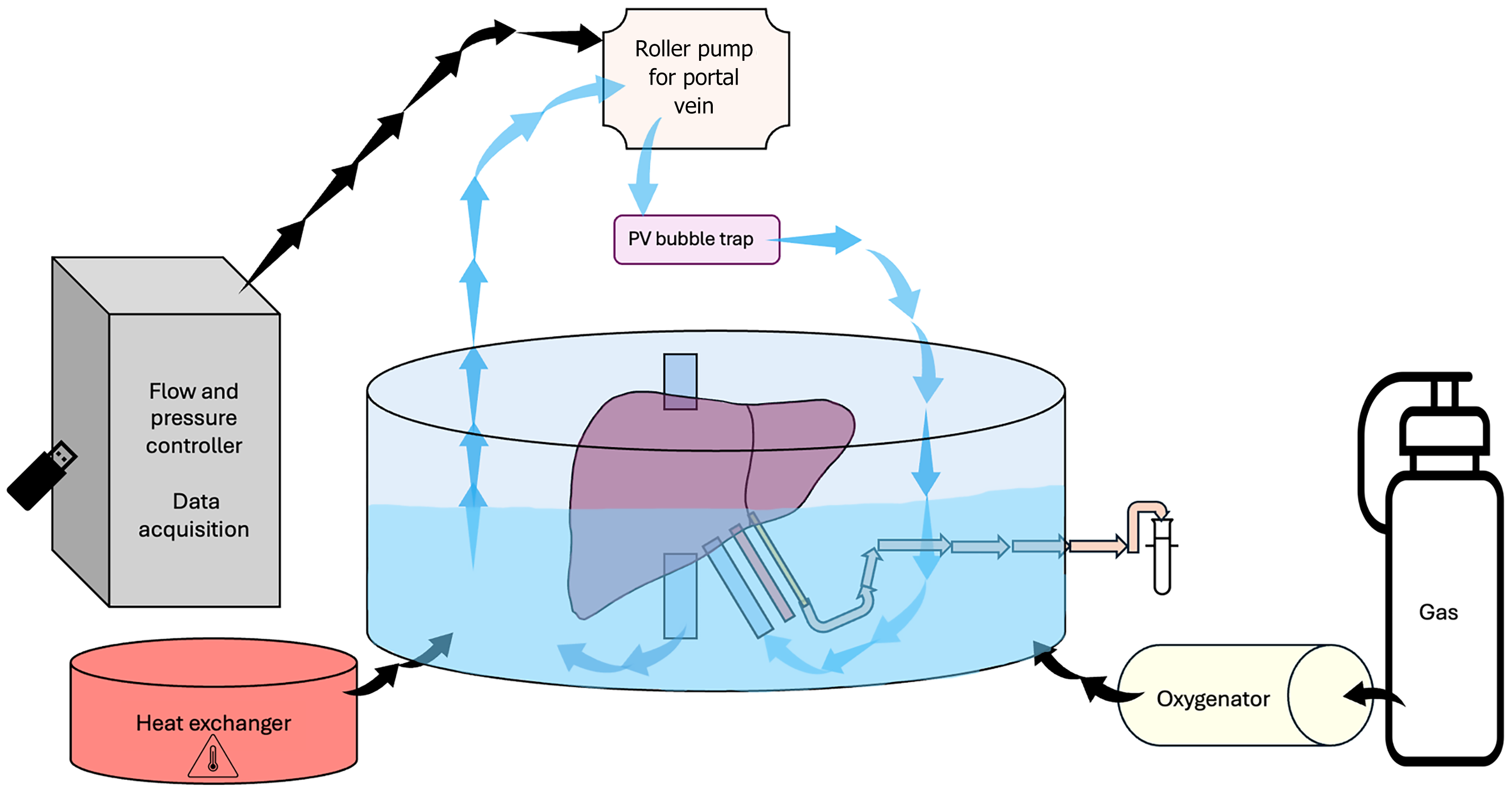
Figure 3 Single-vessel machine perfusion circuit.
The liver is placed inside an organ chamber, which serves as a perfusate reservoir. The perfusate is oxygenated via an oxygenator, and the targeted temperature is achieved via a thermos unit. The perfusate from the organ chamber is then taken up by a roller pump, which is controlled by a flow and pressure controller. After passing through the roller pump, the perfusate goes through a bubble trap and into the portal vein. Upon perfusing the liver, the perfusate exits the liver freely via the vena cava. The common bile duct is cannulated, and the catheter is connected to the tube for bile collection. PV: Portal vein.
- Citation: Bickaite-Bausiene K, Kvietkauskas M, Leber B, Bausys B, Brislinger D, Strupas K, Stiegler P. Small animal ex vivo machine perfusion of the liver: A comprehensive literature review. World J Gastroenterol 2026; 32(9): 111199
- URL: https://www.wjgnet.com/1007-9327/full/v32/i9/111199.htm
- DOI: https://dx.doi.org/10.3748/wjg.v32.i9.111199