
















©The Author(s) 2026.
World J Gastroenterol. Feb 28, 2026; 32(8): 116856
Published online Feb 28, 2026. doi: 10.3748/wjg.v32.i8.116856
Published online Feb 28, 2026. doi: 10.3748/wjg.v32.i8.116856
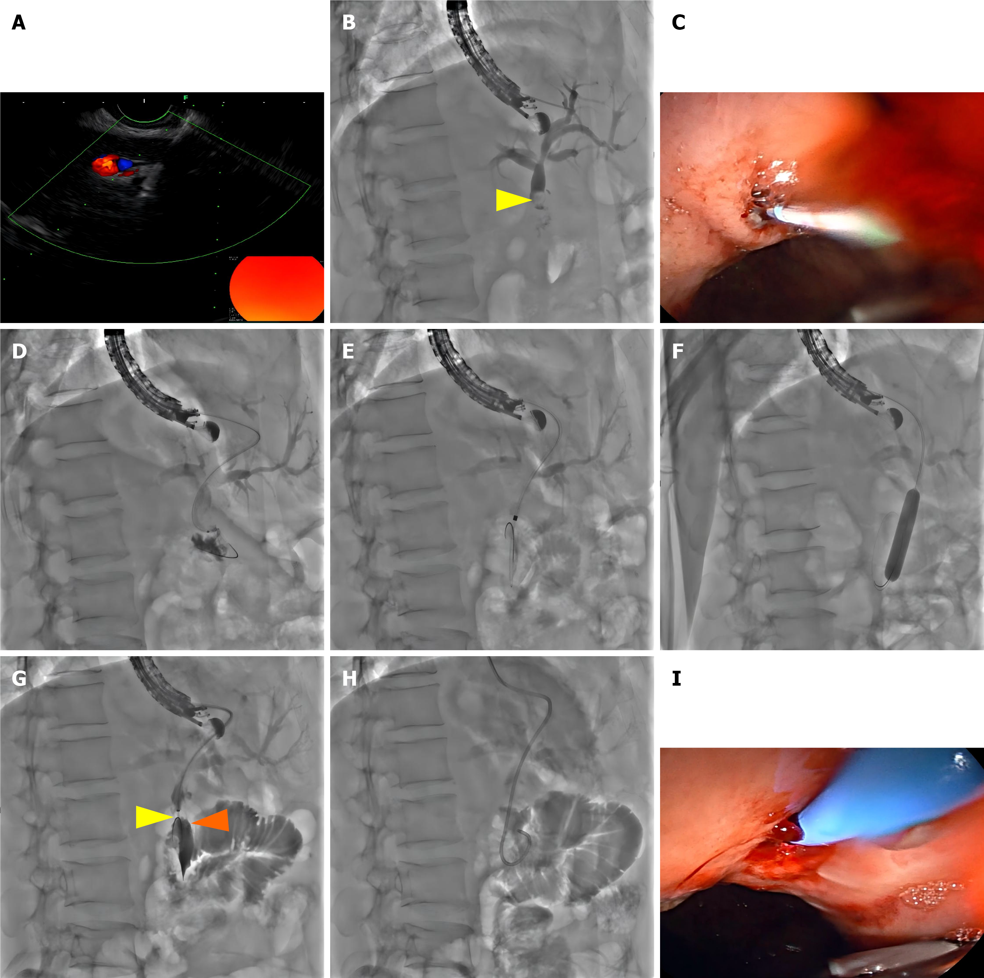
Figure 2 Procedural steps of successful endoscopic ultrasound-guided transhepatic antegrade stone removal.
A: Puncture of a bile duct branch in segment S2 of the liver using a fine-needle aspiration needle; B: Cholangiography confirming mildly dilated intrahepatic bile ducts and a hyperechoic nodule (stone) within the common hepatic duct (yellow arrowheads); C: A needle knife is used to incise the gastric wall and the liver parenchyma to establish a hepatoenteric tract; D: The guidewire is advanced across the biliary-enteric anastomosis into the jejunum; E: Dilation of the created hepatoenteric tract; F: Sequential balloon dilation of the biliary-enteric anastomosis; G: The stone within the common hepatic duct (orange arrowheads) is dislodged and pushed into the jejunum using a retrieval balloon catheter (yellow arrowheads); H and I: Placement of a nasobiliary drainage catheter across the biliary-enteric anastomosis.
- Citation: Li YM, Yasen A, Chen SF, Fan J, Huang XB, Zuo GH, Zheng L. Endoscopic ultrasound-guided transhepatic antegrade stone removal for choledocholithiasis after pancreaticoduodenectomy: A case report and review of literature. World J Gastroenterol 2026; 32(8): 116856
- URL: https://www.wjgnet.com/1007-9327/full/v32/i8/116856.htm
- DOI: https://dx.doi.org/10.3748/wjg.v32.i8.116856