
















©The Author(s) 2026.
World J Gastroenterol. Feb 28, 2026; 32(8): 115654
Published online Feb 28, 2026. doi: 10.3748/wjg.v32.i8.115654
Published online Feb 28, 2026. doi: 10.3748/wjg.v32.i8.115654
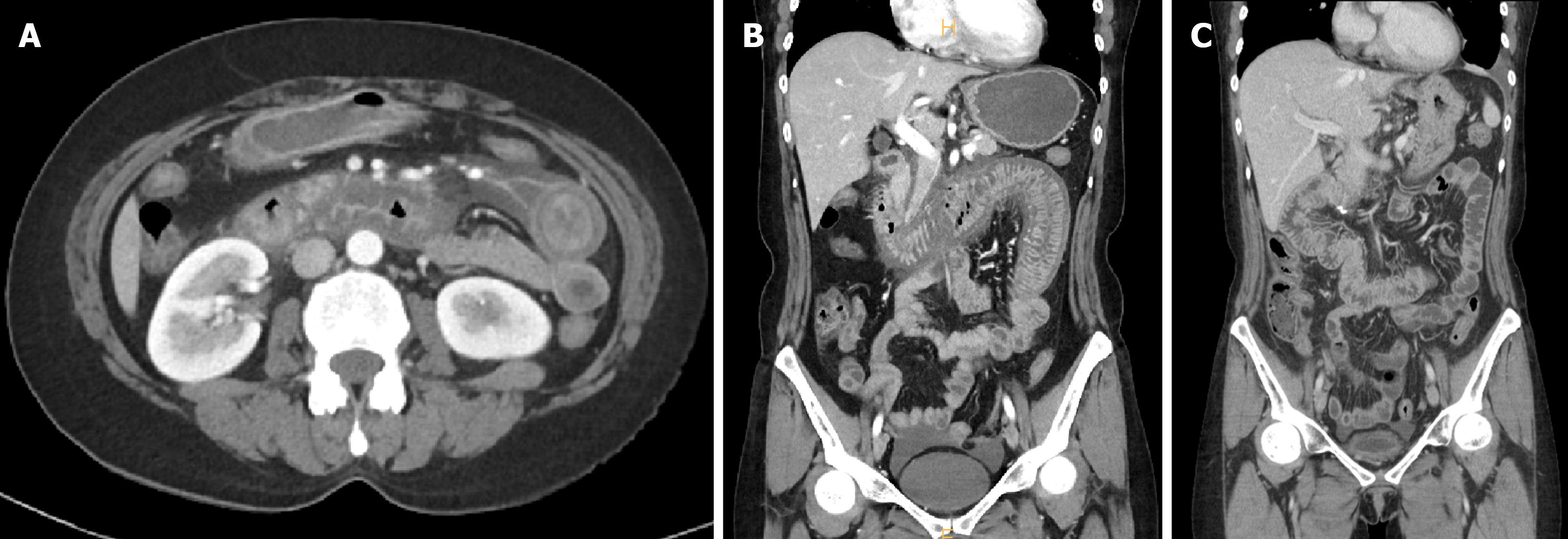
Figure 2 The patient's imaging results.
A: Preoperative contrast-enhanced computed tomography (CT; transverse view) shows duodenal wall thickening and mesenteric congestion; B: Coronal CT view demonstrates venous engorgement and submucosal edema extending from the duodenum to the jejunum; C: Contrast-enhanced abdominal CT at 8 months postoperatively shows an intact anastomosis and no abnormal biliary dilation.
- Citation: Kim YK, Jung HI, Kim H, Bae SH. Ischemic duodenal injury due to systemic lupus erythematosus: A case report. World J Gastroenterol 2026; 32(8): 115654
- URL: https://www.wjgnet.com/1007-9327/full/v32/i8/115654.htm
- DOI: https://dx.doi.org/10.3748/wjg.v32.i8.115654